Gbambele Kone MD, Nino Kvantaliani MD, Brett Cucchiara MD
{"title":"缺血性中风出血性转变中的斑点征。","authors":"Gbambele Kone MD, Nino Kvantaliani MD, Brett Cucchiara MD","doi":"10.1002/ana.26969","DOIUrl":null,"url":null,"abstract":"<p>A 52-year-old man with atrial fibrillation not on anticoagulation presented with 3 days of mild left hemiparesis. He reported taking aspirin 325 mg prior to presentation. Head computed tomography (CT) showed subacute right subcortical infarction (Fig 1A). Several hours later, he developed right gaze deviation and worsened left hemiparesis. Repeat CT showed confluent hemorrhage in the prior infarct; CT angiography performed at the same time showed a spot sign within the hemorrhage (Fig 1B,C). Follow-up imaging demonstrated stable hemorrhage. Hemorrhagic transformation after ischemic stroke is often ascribed to reperfusion of necrotic infarcted tissue with associated blood—brain barrier disruption; ischemia has also been proposed to cause direct vessel injury causing vessel rupture and hemorrhage, though with limited supportive evidence.<span><sup>1, 2</sup></span> The spot sign, as seen here, represents direct evidence of vessel injury with associated contrast extravasation,<span><sup>3</sup></span> supporting the latter as the mechanism in this case. Prior studies have demonstrated that the presence of a spot sign is associated with more than double the risk of both early hematoma expansion and worse functional outcome and mortality in non-traumatic intracerebral hemorrhage.<span><sup>4</sup></span></p><p>G.K., B.C., and N.K. contributed to the conception and design of the study; G.K., B.C., and N.K. contributed to the acquisition and analysis of data; G.K. and B.C. contributed to drafting the manuscript and preparing the figures.</p><p>Nothing to report.</p>","PeriodicalId":127,"journal":{"name":"Annals of Neurology","volume":"96 3","pages":"591-592"},"PeriodicalIF":7.7000,"publicationDate":"2024-05-17","publicationTypes":"Journal Article","fieldsOfStudy":null,"isOpenAccess":false,"openAccessPdf":"https://onlinelibrary.wiley.com/doi/epdf/10.1002/ana.26969","citationCount":"0","resultStr":"{\"title\":\"Spot Sign in Hemorrhagic Transformation of Ischemic Stroke\",\"authors\":\"Gbambele Kone MD, Nino Kvantaliani MD, Brett Cucchiara MD\",\"doi\":\"10.1002/ana.26969\",\"DOIUrl\":null,\"url\":null,\"abstract\":\"<p>A 52-year-old man with atrial fibrillation not on anticoagulation presented with 3 days of mild left hemiparesis. He reported taking aspirin 325 mg prior to presentation. Head computed tomography (CT) showed subacute right subcortical infarction (Fig 1A). Several hours later, he developed right gaze deviation and worsened left hemiparesis. Repeat CT showed confluent hemorrhage in the prior infarct; CT angiography performed at the same time showed a spot sign within the hemorrhage (Fig 1B,C). Follow-up imaging demonstrated stable hemorrhage. Hemorrhagic transformation after ischemic stroke is often ascribed to reperfusion of necrotic infarcted tissue with associated blood—brain barrier disruption; ischemia has also been proposed to cause direct vessel injury causing vessel rupture and hemorrhage, though with limited supportive evidence.<span><sup>1, 2</sup></span> The spot sign, as seen here, represents direct evidence of vessel injury with associated contrast extravasation,<span><sup>3</sup></span> supporting the latter as the mechanism in this case. Prior studies have demonstrated that the presence of a spot sign is associated with more than double the risk of both early hematoma expansion and worse functional outcome and mortality in non-traumatic intracerebral hemorrhage.<span><sup>4</sup></span></p><p>G.K., B.C., and N.K. contributed to the conception and design of the study; G.K., B.C., and N.K. contributed to the acquisition and analysis of data; G.K. and B.C. contributed to drafting the manuscript and preparing the figures.</p><p>Nothing to report.</p>\",\"PeriodicalId\":127,\"journal\":{\"name\":\"Annals of Neurology\",\"volume\":\"96 3\",\"pages\":\"591-592\"},\"PeriodicalIF\":7.7000,\"publicationDate\":\"2024-05-17\",\"publicationTypes\":\"Journal Article\",\"fieldsOfStudy\":null,\"isOpenAccess\":false,\"openAccessPdf\":\"https://onlinelibrary.wiley.com/doi/epdf/10.1002/ana.26969\",\"citationCount\":\"0\",\"resultStr\":null,\"platform\":\"Semanticscholar\",\"paperid\":null,\"PeriodicalName\":\"Annals of Neurology\",\"FirstCategoryId\":\"3\",\"ListUrlMain\":\"https://onlinelibrary.wiley.com/doi/10.1002/ana.26969\",\"RegionNum\":1,\"RegionCategory\":\"医学\",\"ArticlePicture\":[],\"TitleCN\":null,\"AbstractTextCN\":null,\"PMCID\":null,\"EPubDate\":\"\",\"PubModel\":\"\",\"JCR\":\"Q1\",\"JCRName\":\"CLINICAL NEUROLOGY\",\"Score\":null,\"Total\":0}","platform":"Semanticscholar","paperid":null,"PeriodicalName":"Annals of Neurology","FirstCategoryId":"3","ListUrlMain":"https://onlinelibrary.wiley.com/doi/10.1002/ana.26969","RegionNum":1,"RegionCategory":"医学","ArticlePicture":[],"TitleCN":null,"AbstractTextCN":null,"PMCID":null,"EPubDate":"","PubModel":"","JCR":"Q1","JCRName":"CLINICAL NEUROLOGY","Score":null,"Total":0}
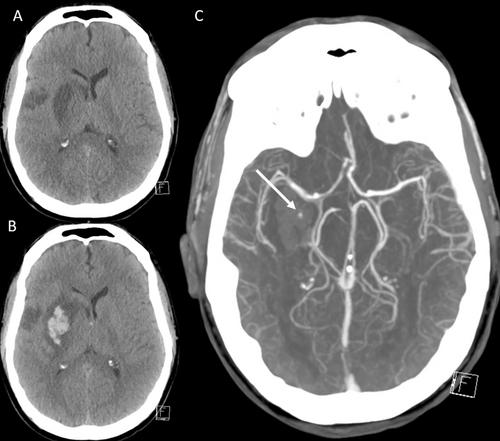
Spot Sign in Hemorrhagic Transformation of Ischemic Stroke
A 52-year-old man with atrial fibrillation not on anticoagulation presented with 3 days of mild left hemiparesis. He reported taking aspirin 325 mg prior to presentation. Head computed tomography (CT) showed subacute right subcortical infarction (Fig 1A). Several hours later, he developed right gaze deviation and worsened left hemiparesis. Repeat CT showed confluent hemorrhage in the prior infarct; CT angiography performed at the same time showed a spot sign within the hemorrhage (Fig 1B,C). Follow-up imaging demonstrated stable hemorrhage. Hemorrhagic transformation after ischemic stroke is often ascribed to reperfusion of necrotic infarcted tissue with associated blood—brain barrier disruption; ischemia has also been proposed to cause direct vessel injury causing vessel rupture and hemorrhage, though with limited supportive evidence.1, 2 The spot sign, as seen here, represents direct evidence of vessel injury with associated contrast extravasation,3 supporting the latter as the mechanism in this case. Prior studies have demonstrated that the presence of a spot sign is associated with more than double the risk of both early hematoma expansion and worse functional outcome and mortality in non-traumatic intracerebral hemorrhage.4
G.K., B.C., and N.K. contributed to the conception and design of the study; G.K., B.C., and N.K. contributed to the acquisition and analysis of data; G.K. and B.C. contributed to drafting the manuscript and preparing the figures.
期刊介绍:
Annals of Neurology publishes original articles with potential for high impact in understanding the pathogenesis, clinical and laboratory features, diagnosis, treatment, outcomes and science underlying diseases of the human nervous system. Articles should ideally be of broad interest to the academic neurological community rather than solely to subspecialists in a particular field. Studies involving experimental model system, including those in cell and organ cultures and animals, of direct translational relevance to the understanding of neurological disease are also encouraged.


 分享
分享
 求助内容:
求助内容: 应助结果提醒方式:
 扫码关注我们
