{"title":"神经退行性疾病中普特曼铁的定量分析:定量易感性绘图技术的荟萃分析。","authors":"Sana Mohammadi, Sadegh Ghaderi, Farzad Fatehi","doi":"10.1007/s11682-024-00895-6","DOIUrl":null,"url":null,"abstract":"<p><p>Quantitative susceptibility mapping (QSM) is an MRI technique that accurately measures iron concentration in brain tissues. This meta-analysis synthesized evidence from 30 studies that used QSM to quantify the iron levels in the putamen. The PRISMA statement was adhered to when conducting the systematic reviews and meta-analyses. We conducted a meta-analysis using a random-effects model, as well as subgroup analyses (disease type, geographic region, field strength, coil, disease type, age, and sex) and sensitivity analysis. A total of 1247 patients and 1035 controls were included in the study. Pooled results showed a standardized mean difference (SMD) of 0.41 (95% CI 0.19 to 0.64), with the strongest effect seen in Alzheimer's disease (AD) at 1.01 (95% CI 0.50 to 1.52). Relapsing-remitting multiple sclerosis (RRMS) also showed increased putaminal iron at 0.37 (95% CI 0.177 to 0.58). No significant differences were observed in Parkinson's disease (PD). No significant differences were found between subgroups based on geographic region, field strength, coil, disease type, age, and sex. The studies revealed significant heterogeneity, with field strength as the primary source, while other factors, such as disease type, location, age, sex, and coil type, may have contributed. The sensitivity analysis showed that these factors did not have a significant influence on the overall results. In summary, this meta-analysis supports abnormalities in putaminal iron content across different diseases with neurodegeneration, especially AD and RRMS, as measured by QSM. This highlights the potential of QSM as an imaging biomarker to better understand disease mechanisms involving disturbances in brain iron homeostasis.</p>","PeriodicalId":9192,"journal":{"name":"Brain Imaging and Behavior","volume":" ","pages":"1239-1255"},"PeriodicalIF":2.4000,"publicationDate":"2024-10-01","publicationTypes":"Journal Article","fieldsOfStudy":null,"isOpenAccess":false,"openAccessPdf":"","citationCount":"0","resultStr":"{\"title\":\"Putamen iron quantification in diseases with neurodegeneration: a meta-analysis of the quantitative susceptibility mapping technique.\",\"authors\":\"Sana Mohammadi, Sadegh Ghaderi, Farzad Fatehi\",\"doi\":\"10.1007/s11682-024-00895-6\",\"DOIUrl\":null,\"url\":null,\"abstract\":\"<p><p>Quantitative susceptibility mapping (QSM) is an MRI technique that accurately measures iron concentration in brain tissues. This meta-analysis synthesized evidence from 30 studies that used QSM to quantify the iron levels in the putamen. The PRISMA statement was adhered to when conducting the systematic reviews and meta-analyses. We conducted a meta-analysis using a random-effects model, as well as subgroup analyses (disease type, geographic region, field strength, coil, disease type, age, and sex) and sensitivity analysis. A total of 1247 patients and 1035 controls were included in the study. Pooled results showed a standardized mean difference (SMD) of 0.41 (95% CI 0.19 to 0.64), with the strongest effect seen in Alzheimer's disease (AD) at 1.01 (95% CI 0.50 to 1.52). Relapsing-remitting multiple sclerosis (RRMS) also showed increased putaminal iron at 0.37 (95% CI 0.177 to 0.58). No significant differences were observed in Parkinson's disease (PD). No significant differences were found between subgroups based on geographic region, field strength, coil, disease type, age, and sex. The studies revealed significant heterogeneity, with field strength as the primary source, while other factors, such as disease type, location, age, sex, and coil type, may have contributed. The sensitivity analysis showed that these factors did not have a significant influence on the overall results. In summary, this meta-analysis supports abnormalities in putaminal iron content across different diseases with neurodegeneration, especially AD and RRMS, as measured by QSM. This highlights the potential of QSM as an imaging biomarker to better understand disease mechanisms involving disturbances in brain iron homeostasis.</p>\",\"PeriodicalId\":9192,\"journal\":{\"name\":\"Brain Imaging and Behavior\",\"volume\":\" \",\"pages\":\"1239-1255\"},\"PeriodicalIF\":2.4000,\"publicationDate\":\"2024-10-01\",\"publicationTypes\":\"Journal Article\",\"fieldsOfStudy\":null,\"isOpenAccess\":false,\"openAccessPdf\":\"\",\"citationCount\":\"0\",\"resultStr\":null,\"platform\":\"Semanticscholar\",\"paperid\":null,\"PeriodicalName\":\"Brain Imaging and Behavior\",\"FirstCategoryId\":\"3\",\"ListUrlMain\":\"https://doi.org/10.1007/s11682-024-00895-6\",\"RegionNum\":3,\"RegionCategory\":\"医学\",\"ArticlePicture\":[],\"TitleCN\":null,\"AbstractTextCN\":null,\"PMCID\":null,\"EPubDate\":\"2024/5/17 0:00:00\",\"PubModel\":\"Epub\",\"JCR\":\"Q2\",\"JCRName\":\"NEUROIMAGING\",\"Score\":null,\"Total\":0}","platform":"Semanticscholar","paperid":null,"PeriodicalName":"Brain Imaging and Behavior","FirstCategoryId":"3","ListUrlMain":"https://doi.org/10.1007/s11682-024-00895-6","RegionNum":3,"RegionCategory":"医学","ArticlePicture":[],"TitleCN":null,"AbstractTextCN":null,"PMCID":null,"EPubDate":"2024/5/17 0:00:00","PubModel":"Epub","JCR":"Q2","JCRName":"NEUROIMAGING","Score":null,"Total":0}
引用次数: 0
摘要
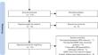
定量易感图(QSM)是一种磁共振成像技术,可精确测量脑组织中的铁浓度。这项荟萃分析综合了 30 项研究的证据,这些研究使用 QSM 量化了普鲁士门的铁含量。在进行系统综述和荟萃分析时,我们遵守了 PRISMA 声明。我们采用随机效应模型进行了荟萃分析,并进行了亚组分析(疾病类型、地理区域、场强、线圈、疾病类型、年龄和性别)和敏感性分析。研究共纳入了 1247 名患者和 1035 名对照组。汇总结果显示,标准化平均差异(SMD)为 0.41(95% CI 0.19 至 0.64),其中阿尔茨海默病(AD)的影响最大,为 1.01(95% CI 0.50 至 1.52)。复发-缓解型多发性硬化症(RRMS)也显示出磷脂膜铁的增加,为 0.37(95% CI 0.177 至 0.58)。在帕金森病(PD)中未观察到明显差异。基于地理区域、场强、线圈、疾病类型、年龄和性别的亚组之间未发现明显差异。这些研究显示出明显的异质性,其中场强是主要原因,而疾病类型、地点、年龄、性别和线圈类型等其他因素可能也有影响。敏感性分析表明,这些因素对总体结果没有显著影响。总之,这项荟萃分析支持通过 QSM 测量神经退行性疾病(尤其是 AD 和 RRMS)中磷脂膜铁含量的异常。这凸显了 QSM 作为成像生物标记物的潜力,有助于更好地了解涉及脑铁平衡紊乱的疾病机制。
Putamen iron quantification in diseases with neurodegeneration: a meta-analysis of the quantitative susceptibility mapping technique.
Quantitative susceptibility mapping (QSM) is an MRI technique that accurately measures iron concentration in brain tissues. This meta-analysis synthesized evidence from 30 studies that used QSM to quantify the iron levels in the putamen. The PRISMA statement was adhered to when conducting the systematic reviews and meta-analyses. We conducted a meta-analysis using a random-effects model, as well as subgroup analyses (disease type, geographic region, field strength, coil, disease type, age, and sex) and sensitivity analysis. A total of 1247 patients and 1035 controls were included in the study. Pooled results showed a standardized mean difference (SMD) of 0.41 (95% CI 0.19 to 0.64), with the strongest effect seen in Alzheimer's disease (AD) at 1.01 (95% CI 0.50 to 1.52). Relapsing-remitting multiple sclerosis (RRMS) also showed increased putaminal iron at 0.37 (95% CI 0.177 to 0.58). No significant differences were observed in Parkinson's disease (PD). No significant differences were found between subgroups based on geographic region, field strength, coil, disease type, age, and sex. The studies revealed significant heterogeneity, with field strength as the primary source, while other factors, such as disease type, location, age, sex, and coil type, may have contributed. The sensitivity analysis showed that these factors did not have a significant influence on the overall results. In summary, this meta-analysis supports abnormalities in putaminal iron content across different diseases with neurodegeneration, especially AD and RRMS, as measured by QSM. This highlights the potential of QSM as an imaging biomarker to better understand disease mechanisms involving disturbances in brain iron homeostasis.
期刊介绍:
Brain Imaging and Behavior is a bi-monthly, peer-reviewed journal, that publishes clinically relevant research using neuroimaging approaches to enhance our understanding of disorders of higher brain function. The journal is targeted at clinicians and researchers in fields concerned with human brain-behavior relationships, such as neuropsychology, psychiatry, neurology, neurosurgery, rehabilitation, and cognitive neuroscience.


 分享
分享
 求助内容:
求助内容: 应助结果提醒方式:
 扫码关注我们
