Tareq Abdel-Alim, Franz Tapia Chaca, Irene M. J. Mathijssen, Clemens M. F. Dirven, Wiro J. Niessen, Eppo B. Wolvius, Marie-Lise C. van Veelen, Gennady V. Roshchupkin
{"title":"利用人工神经网络量化神经颅骨的畸形。","authors":"Tareq Abdel-Alim, Franz Tapia Chaca, Irene M. J. Mathijssen, Clemens M. F. Dirven, Wiro J. Niessen, Eppo B. Wolvius, Marie-Lise C. van Veelen, Gennady V. Roshchupkin","doi":"10.1111/joa.14061","DOIUrl":null,"url":null,"abstract":"<div>\n \n \n <section>\n \n <h3> Background</h3>\n \n <p>Craniosynostosis, a congenital condition characterized by the premature fusion of cranial sutures, necessitates objective methods for evaluating cranial morphology to enhance patient treatment. Current subjective assessments often lead to inconsistent outcomes. This study introduces a novel, quantitative approach to classify craniosynostosis and measure its severity.</p>\n </section>\n \n <section>\n \n <h3> Methods</h3>\n \n <p>An artificial neural network was trained to classify normocephalic, trigonocephalic, and scaphocephalic head shapes based on a publicly available dataset of synthetic 3D head models. Each 3D model was converted into a low-dimensional shape representation based on the distribution of normal vectors, which served as the input for the neural network, ensuring complete patient anonymity and invariance to geometric size and orientation. Explainable AI methods were utilized to highlight significant features when making predictions. Additionally, the Feature Prominence (FP) score was introduced, a novel metric that captures the prominence of distinct shape characteristics associated with a given class. Its relationship with clinical severity scores was examined using the Spearman Rank Correlation Coefficient.</p>\n </section>\n \n <section>\n \n <h3> Results</h3>\n \n <p>The final model achieved excellent test accuracy in classifying the different cranial shapes from their low-dimensional representation. Attention maps indicated that the network's attention was predominantly directed toward the parietal and temporal regions, as well as toward the region signifying vertex depression in scaphocephaly. In trigonocephaly, features around the temples were most pronounced. The FP score showed a strong positive monotonic relationship with clinical severity scores in both scaphocephalic (<i>ρ</i> = 0.83, <i>p</i> < 0.001) and trigonocephalic (<i>ρ</i> = 0.64, <i>p</i> < 0.001) models. Visual assessments further confirmed that as FP values rose, phenotypic severity became increasingly evident.</p>\n </section>\n \n <section>\n \n <h3> Conclusion</h3>\n \n <p>This study presents an innovative and accessible AI-based method for quantifying cranial shape that mitigates the need for adjustments due to age-specific size variations or differences in the spatial orientation of the 3D images, while ensuring complete patient privacy. The proposed FP score strongly correlates with clinical severity scores and has the potential to aid in clinical decision-making and facilitate multi-center collaborations. Future work will focus on validating the model with larger patient datasets and exploring the potential of the FP score for broader applications. The publicly available source code facilitates easy implementation, aiming to advance craniofacial care and research.</p>\n </section>\n </div>","PeriodicalId":14971,"journal":{"name":"Journal of Anatomy","volume":"245 6","pages":"903-913"},"PeriodicalIF":1.9000,"publicationDate":"2024-05-17","publicationTypes":"Journal Article","fieldsOfStudy":null,"isOpenAccess":false,"openAccessPdf":"https://www.ncbi.nlm.nih.gov/pmc/articles/PMC11547242/pdf/","citationCount":"0","resultStr":"{\"title\":\"Quantifying dysmorphologies of the neurocranium using artificial neural networks\",\"authors\":\"Tareq Abdel-Alim, Franz Tapia Chaca, Irene M. J. Mathijssen, Clemens M. F. Dirven, Wiro J. Niessen, Eppo B. Wolvius, Marie-Lise C. van Veelen, Gennady V. Roshchupkin\",\"doi\":\"10.1111/joa.14061\",\"DOIUrl\":null,\"url\":null,\"abstract\":\"<div>\\n \\n \\n <section>\\n \\n <h3> Background</h3>\\n \\n <p>Craniosynostosis, a congenital condition characterized by the premature fusion of cranial sutures, necessitates objective methods for evaluating cranial morphology to enhance patient treatment. Current subjective assessments often lead to inconsistent outcomes. This study introduces a novel, quantitative approach to classify craniosynostosis and measure its severity.</p>\\n </section>\\n \\n <section>\\n \\n <h3> Methods</h3>\\n \\n <p>An artificial neural network was trained to classify normocephalic, trigonocephalic, and scaphocephalic head shapes based on a publicly available dataset of synthetic 3D head models. Each 3D model was converted into a low-dimensional shape representation based on the distribution of normal vectors, which served as the input for the neural network, ensuring complete patient anonymity and invariance to geometric size and orientation. Explainable AI methods were utilized to highlight significant features when making predictions. Additionally, the Feature Prominence (FP) score was introduced, a novel metric that captures the prominence of distinct shape characteristics associated with a given class. Its relationship with clinical severity scores was examined using the Spearman Rank Correlation Coefficient.</p>\\n </section>\\n \\n <section>\\n \\n <h3> Results</h3>\\n \\n <p>The final model achieved excellent test accuracy in classifying the different cranial shapes from their low-dimensional representation. Attention maps indicated that the network's attention was predominantly directed toward the parietal and temporal regions, as well as toward the region signifying vertex depression in scaphocephaly. In trigonocephaly, features around the temples were most pronounced. The FP score showed a strong positive monotonic relationship with clinical severity scores in both scaphocephalic (<i>ρ</i> = 0.83, <i>p</i> < 0.001) and trigonocephalic (<i>ρ</i> = 0.64, <i>p</i> < 0.001) models. Visual assessments further confirmed that as FP values rose, phenotypic severity became increasingly evident.</p>\\n </section>\\n \\n <section>\\n \\n <h3> Conclusion</h3>\\n \\n <p>This study presents an innovative and accessible AI-based method for quantifying cranial shape that mitigates the need for adjustments due to age-specific size variations or differences in the spatial orientation of the 3D images, while ensuring complete patient privacy. The proposed FP score strongly correlates with clinical severity scores and has the potential to aid in clinical decision-making and facilitate multi-center collaborations. Future work will focus on validating the model with larger patient datasets and exploring the potential of the FP score for broader applications. The publicly available source code facilitates easy implementation, aiming to advance craniofacial care and research.</p>\\n </section>\\n </div>\",\"PeriodicalId\":14971,\"journal\":{\"name\":\"Journal of Anatomy\",\"volume\":\"245 6\",\"pages\":\"903-913\"},\"PeriodicalIF\":1.9000,\"publicationDate\":\"2024-05-17\",\"publicationTypes\":\"Journal Article\",\"fieldsOfStudy\":null,\"isOpenAccess\":false,\"openAccessPdf\":\"https://www.ncbi.nlm.nih.gov/pmc/articles/PMC11547242/pdf/\",\"citationCount\":\"0\",\"resultStr\":null,\"platform\":\"Semanticscholar\",\"paperid\":null,\"PeriodicalName\":\"Journal of Anatomy\",\"FirstCategoryId\":\"3\",\"ListUrlMain\":\"https://onlinelibrary.wiley.com/doi/10.1111/joa.14061\",\"RegionNum\":3,\"RegionCategory\":\"医学\",\"ArticlePicture\":[],\"TitleCN\":null,\"AbstractTextCN\":null,\"PMCID\":null,\"EPubDate\":\"\",\"PubModel\":\"\",\"JCR\":\"Q2\",\"JCRName\":\"ANATOMY & MORPHOLOGY\",\"Score\":null,\"Total\":0}","platform":"Semanticscholar","paperid":null,"PeriodicalName":"Journal of Anatomy","FirstCategoryId":"3","ListUrlMain":"https://onlinelibrary.wiley.com/doi/10.1111/joa.14061","RegionNum":3,"RegionCategory":"医学","ArticlePicture":[],"TitleCN":null,"AbstractTextCN":null,"PMCID":null,"EPubDate":"","PubModel":"","JCR":"Q2","JCRName":"ANATOMY & MORPHOLOGY","Score":null,"Total":0}
Quantifying dysmorphologies of the neurocranium using artificial neural networks
Background
Craniosynostosis, a congenital condition characterized by the premature fusion of cranial sutures, necessitates objective methods for evaluating cranial morphology to enhance patient treatment. Current subjective assessments often lead to inconsistent outcomes. This study introduces a novel, quantitative approach to classify craniosynostosis and measure its severity.
Methods
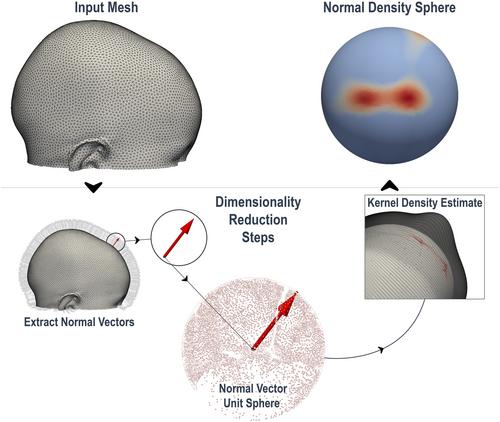
An artificial neural network was trained to classify normocephalic, trigonocephalic, and scaphocephalic head shapes based on a publicly available dataset of synthetic 3D head models. Each 3D model was converted into a low-dimensional shape representation based on the distribution of normal vectors, which served as the input for the neural network, ensuring complete patient anonymity and invariance to geometric size and orientation. Explainable AI methods were utilized to highlight significant features when making predictions. Additionally, the Feature Prominence (FP) score was introduced, a novel metric that captures the prominence of distinct shape characteristics associated with a given class. Its relationship with clinical severity scores was examined using the Spearman Rank Correlation Coefficient.
Results
The final model achieved excellent test accuracy in classifying the different cranial shapes from their low-dimensional representation. Attention maps indicated that the network's attention was predominantly directed toward the parietal and temporal regions, as well as toward the region signifying vertex depression in scaphocephaly. In trigonocephaly, features around the temples were most pronounced. The FP score showed a strong positive monotonic relationship with clinical severity scores in both scaphocephalic (ρ = 0.83, p < 0.001) and trigonocephalic (ρ = 0.64, p < 0.001) models. Visual assessments further confirmed that as FP values rose, phenotypic severity became increasingly evident.
Conclusion
This study presents an innovative and accessible AI-based method for quantifying cranial shape that mitigates the need for adjustments due to age-specific size variations or differences in the spatial orientation of the 3D images, while ensuring complete patient privacy. The proposed FP score strongly correlates with clinical severity scores and has the potential to aid in clinical decision-making and facilitate multi-center collaborations. Future work will focus on validating the model with larger patient datasets and exploring the potential of the FP score for broader applications. The publicly available source code facilitates easy implementation, aiming to advance craniofacial care and research.
期刊介绍:
Journal of Anatomy is an international peer-reviewed journal sponsored by the Anatomical Society. The journal publishes original papers, invited review articles and book reviews. Its main focus is to understand anatomy through an analysis of structure, function, development and evolution. Priority will be given to studies of that clearly articulate their relevance to the anatomical community. Focal areas include: experimental studies, contributions based on molecular and cell biology and on the application of modern imaging techniques and papers with novel methods or synthetic perspective on an anatomical system.
Studies that are essentially descriptive anatomy are appropriate only if they communicate clearly a broader functional or evolutionary significance. You must clearly state the broader implications of your work in the abstract.
We particularly welcome submissions in the following areas:
Cell biology and tissue architecture
Comparative functional morphology
Developmental biology
Evolutionary developmental biology
Evolutionary morphology
Functional human anatomy
Integrative vertebrate paleontology
Methodological innovations in anatomical research
Musculoskeletal system
Neuroanatomy and neurodegeneration
Significant advances in anatomical education.


 分享
分享
 求助内容:
求助内容: 应助结果提醒方式:
 扫码关注我们
