Jie Sun, Chen Yao, Wanxin Luo, Xingyu Ge, Wenjie Zheng, Chi Sun, Yafeng Zhang
{"title":"内皮细胞衍生的外泌体通过激活 PI3K/Akt/Bcl-2 通路抑制成骨细胞凋亡和类固醇诱导的股骨头坏死进程","authors":"Jie Sun, Chen Yao, Wanxin Luo, Xingyu Ge, Wenjie Zheng, Chi Sun, Yafeng Zhang","doi":"10.1155/2024/3870988","DOIUrl":null,"url":null,"abstract":"<div>\n <p>The aim of the study was to investigate the therapeutic potential of exosomes secreted by endothelial cells (EC-exos) on steroid-induced osteonecrosis of femoral head (SNFH). First, we successfully obtained EC-exos through differential centrifugation. Then, the effects of EC-exos on mouse embryo osteoblast precursor (MC3T3-E1) cells under high concentration of dexamethasone (Dex) were analysed <i>in vitro</i>, which included cell migration, viability, and apoptosis. <i>In vivo</i>, a SNFH rat model was successfully established and treated with EC-exos. Micro-computed tomography (micro-CT) and haematoxylin and eosin (H&E) were used to observe femoral trabeculae. Our <i>in vitro</i> results showed that EC-exos improved cell viability and migration of osteoblasts and reduced the apoptotic effect of high concentration of Dex on osteoblasts <i>in vitro</i>. Phosphoinositide 3-kinase (PI3K)/Akt/Bcl-2 signalling pathway was activated in MC3T3-E1 cells under the response to EC-exos. <i>In vivo</i>, increased bone volume per tissue volume (BV/TV) (<i>p</i> = 0.031), trabecular thickness (Tb.Th) (<i>p</i> = 0.020), and decreased separation (Tb.Sp) (<i>p</i> = 0.040) were observed in SNFH rats treated with EC-exos. H&E staining revealed fewer empty lacunae and pyknotic osteocytes in trabeculae. The expression of Bcl-2 and Akt in EC-exos group was significantly increased in trabeculae tissue. Overall, our finding indicated that EC-exos could attenuate SNFH by inhibiting osteoblast apoptosis via the PI3K/Akt/Bcl-2 pathway.</p>\n </div>","PeriodicalId":202,"journal":{"name":"Journal of Tissue Engineering and Regenerative Medicine","volume":"2024 1","pages":""},"PeriodicalIF":2.6000,"publicationDate":"2024-05-10","publicationTypes":"Journal Article","fieldsOfStudy":null,"isOpenAccess":false,"openAccessPdf":"https://onlinelibrary.wiley.com/doi/epdf/10.1155/2024/3870988","citationCount":"0","resultStr":"{\"title\":\"Endothelial Cell-Derived Exosomes Inhibit Osteoblast Apoptosis and Steroid-Induced Necrosis of Femoral Head Progression by Activating the PI3K/Akt/Bcl-2 Pathway\",\"authors\":\"Jie Sun, Chen Yao, Wanxin Luo, Xingyu Ge, Wenjie Zheng, Chi Sun, Yafeng Zhang\",\"doi\":\"10.1155/2024/3870988\",\"DOIUrl\":null,\"url\":null,\"abstract\":\"<div>\\n <p>The aim of the study was to investigate the therapeutic potential of exosomes secreted by endothelial cells (EC-exos) on steroid-induced osteonecrosis of femoral head (SNFH). First, we successfully obtained EC-exos through differential centrifugation. Then, the effects of EC-exos on mouse embryo osteoblast precursor (MC3T3-E1) cells under high concentration of dexamethasone (Dex) were analysed <i>in vitro</i>, which included cell migration, viability, and apoptosis. <i>In vivo</i>, a SNFH rat model was successfully established and treated with EC-exos. Micro-computed tomography (micro-CT) and haematoxylin and eosin (H&E) were used to observe femoral trabeculae. Our <i>in vitro</i> results showed that EC-exos improved cell viability and migration of osteoblasts and reduced the apoptotic effect of high concentration of Dex on osteoblasts <i>in vitro</i>. Phosphoinositide 3-kinase (PI3K)/Akt/Bcl-2 signalling pathway was activated in MC3T3-E1 cells under the response to EC-exos. <i>In vivo</i>, increased bone volume per tissue volume (BV/TV) (<i>p</i> = 0.031), trabecular thickness (Tb.Th) (<i>p</i> = 0.020), and decreased separation (Tb.Sp) (<i>p</i> = 0.040) were observed in SNFH rats treated with EC-exos. H&E staining revealed fewer empty lacunae and pyknotic osteocytes in trabeculae. The expression of Bcl-2 and Akt in EC-exos group was significantly increased in trabeculae tissue. Overall, our finding indicated that EC-exos could attenuate SNFH by inhibiting osteoblast apoptosis via the PI3K/Akt/Bcl-2 pathway.</p>\\n </div>\",\"PeriodicalId\":202,\"journal\":{\"name\":\"Journal of Tissue Engineering and Regenerative Medicine\",\"volume\":\"2024 1\",\"pages\":\"\"},\"PeriodicalIF\":2.6000,\"publicationDate\":\"2024-05-10\",\"publicationTypes\":\"Journal Article\",\"fieldsOfStudy\":null,\"isOpenAccess\":false,\"openAccessPdf\":\"https://onlinelibrary.wiley.com/doi/epdf/10.1155/2024/3870988\",\"citationCount\":\"0\",\"resultStr\":null,\"platform\":\"Semanticscholar\",\"paperid\":null,\"PeriodicalName\":\"Journal of Tissue Engineering and Regenerative Medicine\",\"FirstCategoryId\":\"5\",\"ListUrlMain\":\"https://onlinelibrary.wiley.com/doi/10.1155/2024/3870988\",\"RegionNum\":3,\"RegionCategory\":\"生物学\",\"ArticlePicture\":[],\"TitleCN\":null,\"AbstractTextCN\":null,\"PMCID\":null,\"EPubDate\":\"\",\"PubModel\":\"\",\"JCR\":\"Q2\",\"JCRName\":\"BIOTECHNOLOGY & APPLIED MICROBIOLOGY\",\"Score\":null,\"Total\":0}","platform":"Semanticscholar","paperid":null,"PeriodicalName":"Journal of Tissue Engineering and Regenerative Medicine","FirstCategoryId":"5","ListUrlMain":"https://onlinelibrary.wiley.com/doi/10.1155/2024/3870988","RegionNum":3,"RegionCategory":"生物学","ArticlePicture":[],"TitleCN":null,"AbstractTextCN":null,"PMCID":null,"EPubDate":"","PubModel":"","JCR":"Q2","JCRName":"BIOTECHNOLOGY & APPLIED MICROBIOLOGY","Score":null,"Total":0}
Endothelial Cell-Derived Exosomes Inhibit Osteoblast Apoptosis and Steroid-Induced Necrosis of Femoral Head Progression by Activating the PI3K/Akt/Bcl-2 Pathway
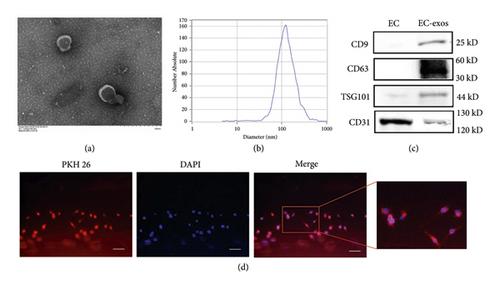
The aim of the study was to investigate the therapeutic potential of exosomes secreted by endothelial cells (EC-exos) on steroid-induced osteonecrosis of femoral head (SNFH). First, we successfully obtained EC-exos through differential centrifugation. Then, the effects of EC-exos on mouse embryo osteoblast precursor (MC3T3-E1) cells under high concentration of dexamethasone (Dex) were analysed in vitro, which included cell migration, viability, and apoptosis. In vivo, a SNFH rat model was successfully established and treated with EC-exos. Micro-computed tomography (micro-CT) and haematoxylin and eosin (H&E) were used to observe femoral trabeculae. Our in vitro results showed that EC-exos improved cell viability and migration of osteoblasts and reduced the apoptotic effect of high concentration of Dex on osteoblasts in vitro. Phosphoinositide 3-kinase (PI3K)/Akt/Bcl-2 signalling pathway was activated in MC3T3-E1 cells under the response to EC-exos. In vivo, increased bone volume per tissue volume (BV/TV) (p = 0.031), trabecular thickness (Tb.Th) (p = 0.020), and decreased separation (Tb.Sp) (p = 0.040) were observed in SNFH rats treated with EC-exos. H&E staining revealed fewer empty lacunae and pyknotic osteocytes in trabeculae. The expression of Bcl-2 and Akt in EC-exos group was significantly increased in trabeculae tissue. Overall, our finding indicated that EC-exos could attenuate SNFH by inhibiting osteoblast apoptosis via the PI3K/Akt/Bcl-2 pathway.
期刊介绍:
Journal of Tissue Engineering and Regenerative Medicine publishes rapidly and rigorously peer-reviewed research papers, reviews, clinical case reports, perspectives, and short communications on topics relevant to the development of therapeutic approaches which combine stem or progenitor cells, biomaterials and scaffolds, growth factors and other bioactive agents, and their respective constructs. All papers should deal with research that has a direct or potential impact on the development of novel clinical approaches for the regeneration or repair of tissues and organs.
The journal is multidisciplinary, covering the combination of the principles of life sciences and engineering in efforts to advance medicine and clinical strategies. The journal focuses on the use of cells, materials, and biochemical/mechanical factors in the development of biological functional substitutes that restore, maintain, or improve tissue or organ function. The journal publishes research on any tissue or organ and covers all key aspects of the field, including the development of new biomaterials and processing of scaffolds; the use of different types of cells (mainly stem and progenitor cells) and their culture in specific bioreactors; studies in relevant animal models; and clinical trials in human patients performed under strict regulatory and ethical frameworks. Manuscripts describing the use of advanced methods for the characterization of engineered tissues are also of special interest to the journal readership.


 分享
分享
 求助内容:
求助内容: 应助结果提醒方式:
 扫码关注我们
