Giuseppe Pontillo, Mario Tranfa, Alessandra Scaravilli, Serena Monti, Ivana Capuano, Eleonora Riccio, Manuela Rizzo, Arturo Brunetti, Giuseppe Palma, Antonio Pisani, Sirio Cocozza
{"title":"利用核磁共振弛豫测量法在法布里病患者脑内展示球形糖基甘油酰胺的积累。","authors":"Giuseppe Pontillo, Mario Tranfa, Alessandra Scaravilli, Serena Monti, Ivana Capuano, Eleonora Riccio, Manuela Rizzo, Arturo Brunetti, Giuseppe Palma, Antonio Pisani, Sirio Cocozza","doi":"10.1007/s00234-024-03380-5","DOIUrl":null,"url":null,"abstract":"<p><strong>Purpose: </strong>How to measure brain globotriaosylceramide (Gb3) accumulation in Fabry Disease (FD) patients in-vivo is still an open challenge. The objective of this study is to provide a quantitative, non-invasive demonstration of this phenomenon using quantitative MRI (qMRI).</p><p><strong>Methods: </strong>In this retrospective, monocentric cross-sectional study conducted from November 2015 to July 2018, FD patients and healthy controls (HC) underwent an MRI scan with a relaxometry protocol to compute longitudinal relaxation rate (R1) maps to evaluate gray (GM) and white matter (WM) lipid accumulation. In a subgroup of 22 FD patients, clinical (FAbry STabilization indEX -FASTEX- score) and biochemical (residual α-galactosidase activity) variables were correlated with MRI data. Quantitative maps were analyzed at both global (\"bulk\" analysis) and regional (\"voxel-wise\" analysis) levels.</p><p><strong>Results: </strong>Data were obtained from 42 FD patients (mean age = 42.4 ± 12.9, M/F = 16/26) and 49 HC (mean age = 42.3 ± 16.3, M/F = 28/21). Compared to HC, FD patients showed a widespread increase in R1 values encompassing both GM (p<sub>FWE</sub> = 0.02) and WM (p<sub>FWE</sub> = 0.02) structures. While no correlations were found between increased R1 values and FASTEX score, a significant negative correlation emerged between residual enzymatic activity levels and R1 values in GM (r = -0.57, p = 0.008) and WM (r = -0.49, p = 0.03).</p><p><strong>Conclusions: </strong>We demonstrated the feasibility and clinical relevance of non-invasively assessing cerebral Gb3 accumulation in FD using MRI. R1 mapping might be used as an in-vivo quantitative neuroimaging biomarker in FD patients.</p>","PeriodicalId":19422,"journal":{"name":"Neuroradiology","volume":" ","pages":"1593-1601"},"PeriodicalIF":2.8000,"publicationDate":"2024-09-01","publicationTypes":"Journal Article","fieldsOfStudy":null,"isOpenAccess":false,"openAccessPdf":"https://www.ncbi.nlm.nih.gov/pmc/articles/PMC11322198/pdf/","citationCount":"0","resultStr":"{\"title\":\"In vivo demonstration of globotriaosylceramide brain accumulation in Fabry Disease using MR Relaxometry.\",\"authors\":\"Giuseppe Pontillo, Mario Tranfa, Alessandra Scaravilli, Serena Monti, Ivana Capuano, Eleonora Riccio, Manuela Rizzo, Arturo Brunetti, Giuseppe Palma, Antonio Pisani, Sirio Cocozza\",\"doi\":\"10.1007/s00234-024-03380-5\",\"DOIUrl\":null,\"url\":null,\"abstract\":\"<p><strong>Purpose: </strong>How to measure brain globotriaosylceramide (Gb3) accumulation in Fabry Disease (FD) patients in-vivo is still an open challenge. The objective of this study is to provide a quantitative, non-invasive demonstration of this phenomenon using quantitative MRI (qMRI).</p><p><strong>Methods: </strong>In this retrospective, monocentric cross-sectional study conducted from November 2015 to July 2018, FD patients and healthy controls (HC) underwent an MRI scan with a relaxometry protocol to compute longitudinal relaxation rate (R1) maps to evaluate gray (GM) and white matter (WM) lipid accumulation. In a subgroup of 22 FD patients, clinical (FAbry STabilization indEX -FASTEX- score) and biochemical (residual α-galactosidase activity) variables were correlated with MRI data. Quantitative maps were analyzed at both global (\\\"bulk\\\" analysis) and regional (\\\"voxel-wise\\\" analysis) levels.</p><p><strong>Results: </strong>Data were obtained from 42 FD patients (mean age = 42.4 ± 12.9, M/F = 16/26) and 49 HC (mean age = 42.3 ± 16.3, M/F = 28/21). Compared to HC, FD patients showed a widespread increase in R1 values encompassing both GM (p<sub>FWE</sub> = 0.02) and WM (p<sub>FWE</sub> = 0.02) structures. While no correlations were found between increased R1 values and FASTEX score, a significant negative correlation emerged between residual enzymatic activity levels and R1 values in GM (r = -0.57, p = 0.008) and WM (r = -0.49, p = 0.03).</p><p><strong>Conclusions: </strong>We demonstrated the feasibility and clinical relevance of non-invasively assessing cerebral Gb3 accumulation in FD using MRI. R1 mapping might be used as an in-vivo quantitative neuroimaging biomarker in FD patients.</p>\",\"PeriodicalId\":19422,\"journal\":{\"name\":\"Neuroradiology\",\"volume\":\" \",\"pages\":\"1593-1601\"},\"PeriodicalIF\":2.8000,\"publicationDate\":\"2024-09-01\",\"publicationTypes\":\"Journal Article\",\"fieldsOfStudy\":null,\"isOpenAccess\":false,\"openAccessPdf\":\"https://www.ncbi.nlm.nih.gov/pmc/articles/PMC11322198/pdf/\",\"citationCount\":\"0\",\"resultStr\":null,\"platform\":\"Semanticscholar\",\"paperid\":null,\"PeriodicalName\":\"Neuroradiology\",\"FirstCategoryId\":\"3\",\"ListUrlMain\":\"https://doi.org/10.1007/s00234-024-03380-5\",\"RegionNum\":3,\"RegionCategory\":\"医学\",\"ArticlePicture\":[],\"TitleCN\":null,\"AbstractTextCN\":null,\"PMCID\":null,\"EPubDate\":\"2024/5/21 0:00:00\",\"PubModel\":\"Epub\",\"JCR\":\"Q2\",\"JCRName\":\"CLINICAL NEUROLOGY\",\"Score\":null,\"Total\":0}","platform":"Semanticscholar","paperid":null,"PeriodicalName":"Neuroradiology","FirstCategoryId":"3","ListUrlMain":"https://doi.org/10.1007/s00234-024-03380-5","RegionNum":3,"RegionCategory":"医学","ArticlePicture":[],"TitleCN":null,"AbstractTextCN":null,"PMCID":null,"EPubDate":"2024/5/21 0:00:00","PubModel":"Epub","JCR":"Q2","JCRName":"CLINICAL NEUROLOGY","Score":null,"Total":0}
In vivo demonstration of globotriaosylceramide brain accumulation in Fabry Disease using MR Relaxometry.
Purpose: How to measure brain globotriaosylceramide (Gb3) accumulation in Fabry Disease (FD) patients in-vivo is still an open challenge. The objective of this study is to provide a quantitative, non-invasive demonstration of this phenomenon using quantitative MRI (qMRI).
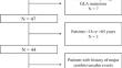
Methods: In this retrospective, monocentric cross-sectional study conducted from November 2015 to July 2018, FD patients and healthy controls (HC) underwent an MRI scan with a relaxometry protocol to compute longitudinal relaxation rate (R1) maps to evaluate gray (GM) and white matter (WM) lipid accumulation. In a subgroup of 22 FD patients, clinical (FAbry STabilization indEX -FASTEX- score) and biochemical (residual α-galactosidase activity) variables were correlated with MRI data. Quantitative maps were analyzed at both global ("bulk" analysis) and regional ("voxel-wise" analysis) levels.
Results: Data were obtained from 42 FD patients (mean age = 42.4 ± 12.9, M/F = 16/26) and 49 HC (mean age = 42.3 ± 16.3, M/F = 28/21). Compared to HC, FD patients showed a widespread increase in R1 values encompassing both GM (pFWE = 0.02) and WM (pFWE = 0.02) structures. While no correlations were found between increased R1 values and FASTEX score, a significant negative correlation emerged between residual enzymatic activity levels and R1 values in GM (r = -0.57, p = 0.008) and WM (r = -0.49, p = 0.03).
Conclusions: We demonstrated the feasibility and clinical relevance of non-invasively assessing cerebral Gb3 accumulation in FD using MRI. R1 mapping might be used as an in-vivo quantitative neuroimaging biomarker in FD patients.
期刊介绍:
Neuroradiology aims to provide state-of-the-art medical and scientific information in the fields of Neuroradiology, Neurosciences, Neurology, Psychiatry, Neurosurgery, and related medical specialities. Neuroradiology as the official Journal of the European Society of Neuroradiology receives submissions from all parts of the world and publishes peer-reviewed original research, comprehensive reviews, educational papers, opinion papers, and short reports on exceptional clinical observations and new technical developments in the field of Neuroimaging and Neurointervention. The journal has subsections for Diagnostic and Interventional Neuroradiology, Advanced Neuroimaging, Paediatric Neuroradiology, Head-Neck-ENT Radiology, Spine Neuroradiology, and for submissions from Japan. Neuroradiology aims to provide new knowledge about and insights into the function and pathology of the human nervous system that may help to better diagnose and treat nervous system diseases. Neuroradiology is a member of the Committee on Publication Ethics (COPE) and follows the COPE core practices. Neuroradiology prefers articles that are free of bias, self-critical regarding limitations, transparent and clear in describing study participants, methods, and statistics, and short in presenting results. Before peer-review all submissions are automatically checked by iThenticate to assess for potential overlap in prior publication.


 分享
分享
 求助内容:
求助内容: 应助结果提醒方式:
 扫码关注我们
