Weiqi Xu, Xinfeng Fei, Zeyu Cui, Dikang Pan, Yan Liu, Te Liu
{"title":"小鼠羊水间充质干细胞外泌体驱动的DNMT1通过诱导角膜上皮细胞microRNA-33启动子DNA高甲基化修饰改善角膜冷冻损伤。","authors":"Weiqi Xu, Xinfeng Fei, Zeyu Cui, Dikang Pan, Yan Liu, Te Liu","doi":"10.1007/s13577-024-01082-x","DOIUrl":null,"url":null,"abstract":"<p><p>Severe corneal cryoinjury can cause permanent corneal swelling and bullous keratopathy, one of the main reason for loss of sight. Mouse amniotic fluid mesenchymal stem cells (mAF-MSCs) can repair corneal damage caused by freezing; however, whether the exosomes derived from mAF-MSCs have the same repair effect is unknown. In this study, the mAF-MSC-exosomes were transplanted into the eyeballs of corneal cryoinjured mice. Histopathological examination showed that the mAF-MSC-exosomes improved the corneal structure and status of corneal epithelial cells in corneal cryoinjured mice. RRBS-sequencing showed that compared with the control group, four genes (Rpl13-ps6, miR-33, Hymai, and Plagl1), underwent DNA hypermethylation modification after mAF-MSC-exosomes treatment. The result of FISH indicated that miR-33-3p hybridization signals were enhanced in corneal epithelial cells from mice treated with mAF-MSC-exosomes. Semi-quantitative PCR and western blotting indicated that mAF-MSC-exosomes contained high levels of DNMT1 mRNA and protein. Additionally, luciferase report assays indicated that miR-33-3p overexpression in NIH-3T3 mouse embryonic fibroblast cells inhibited the activity of luciferase carrying a sequence from the 3' untranslated region of Bcl6. Moreover, BCL6 mRNA and protein levels in corneal tissues from mice treated with mAF-MSC-exosomes were higher than those in the control group. Therefore, our results suggested that mAF-MSC-exosomes could repair corneal cryoinjury by releasing DNMT1, which induced hypermethylation of the miR-33 promoter in corneal epithelial cells. Consequent downregulated miR-33 transcription upregulated Bcl6 expression, ultimately achieving the repair of corneal cryoinjury in mice.</p>","PeriodicalId":49194,"journal":{"name":"Human Cell","volume":" ","pages":"1091-1106"},"PeriodicalIF":3.1000,"publicationDate":"2024-07-01","publicationTypes":"Journal Article","fieldsOfStudy":null,"isOpenAccess":false,"openAccessPdf":"","citationCount":"0","resultStr":"{\"title\":\"DNMT1 driven by mouse amniotic fluid mesenchymal stem cell exosomes improved corneal cryoinjury via inducing microRNA-33 promoter DNA hypermethylation modification in corneal epithelium cells.\",\"authors\":\"Weiqi Xu, Xinfeng Fei, Zeyu Cui, Dikang Pan, Yan Liu, Te Liu\",\"doi\":\"10.1007/s13577-024-01082-x\",\"DOIUrl\":null,\"url\":null,\"abstract\":\"<p><p>Severe corneal cryoinjury can cause permanent corneal swelling and bullous keratopathy, one of the main reason for loss of sight. Mouse amniotic fluid mesenchymal stem cells (mAF-MSCs) can repair corneal damage caused by freezing; however, whether the exosomes derived from mAF-MSCs have the same repair effect is unknown. In this study, the mAF-MSC-exosomes were transplanted into the eyeballs of corneal cryoinjured mice. Histopathological examination showed that the mAF-MSC-exosomes improved the corneal structure and status of corneal epithelial cells in corneal cryoinjured mice. RRBS-sequencing showed that compared with the control group, four genes (Rpl13-ps6, miR-33, Hymai, and Plagl1), underwent DNA hypermethylation modification after mAF-MSC-exosomes treatment. The result of FISH indicated that miR-33-3p hybridization signals were enhanced in corneal epithelial cells from mice treated with mAF-MSC-exosomes. Semi-quantitative PCR and western blotting indicated that mAF-MSC-exosomes contained high levels of DNMT1 mRNA and protein. Additionally, luciferase report assays indicated that miR-33-3p overexpression in NIH-3T3 mouse embryonic fibroblast cells inhibited the activity of luciferase carrying a sequence from the 3' untranslated region of Bcl6. Moreover, BCL6 mRNA and protein levels in corneal tissues from mice treated with mAF-MSC-exosomes were higher than those in the control group. Therefore, our results suggested that mAF-MSC-exosomes could repair corneal cryoinjury by releasing DNMT1, which induced hypermethylation of the miR-33 promoter in corneal epithelial cells. Consequent downregulated miR-33 transcription upregulated Bcl6 expression, ultimately achieving the repair of corneal cryoinjury in mice.</p>\",\"PeriodicalId\":49194,\"journal\":{\"name\":\"Human Cell\",\"volume\":\" \",\"pages\":\"1091-1106\"},\"PeriodicalIF\":3.1000,\"publicationDate\":\"2024-07-01\",\"publicationTypes\":\"Journal Article\",\"fieldsOfStudy\":null,\"isOpenAccess\":false,\"openAccessPdf\":\"\",\"citationCount\":\"0\",\"resultStr\":null,\"platform\":\"Semanticscholar\",\"paperid\":null,\"PeriodicalName\":\"Human Cell\",\"FirstCategoryId\":\"99\",\"ListUrlMain\":\"https://doi.org/10.1007/s13577-024-01082-x\",\"RegionNum\":3,\"RegionCategory\":\"生物学\",\"ArticlePicture\":[],\"TitleCN\":null,\"AbstractTextCN\":null,\"PMCID\":null,\"EPubDate\":\"2024/5/23 0:00:00\",\"PubModel\":\"Epub\",\"JCR\":\"Q3\",\"JCRName\":\"CELL BIOLOGY\",\"Score\":null,\"Total\":0}","platform":"Semanticscholar","paperid":null,"PeriodicalName":"Human Cell","FirstCategoryId":"99","ListUrlMain":"https://doi.org/10.1007/s13577-024-01082-x","RegionNum":3,"RegionCategory":"生物学","ArticlePicture":[],"TitleCN":null,"AbstractTextCN":null,"PMCID":null,"EPubDate":"2024/5/23 0:00:00","PubModel":"Epub","JCR":"Q3","JCRName":"CELL BIOLOGY","Score":null,"Total":0}
DNMT1 driven by mouse amniotic fluid mesenchymal stem cell exosomes improved corneal cryoinjury via inducing microRNA-33 promoter DNA hypermethylation modification in corneal epithelium cells.
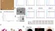
Severe corneal cryoinjury can cause permanent corneal swelling and bullous keratopathy, one of the main reason for loss of sight. Mouse amniotic fluid mesenchymal stem cells (mAF-MSCs) can repair corneal damage caused by freezing; however, whether the exosomes derived from mAF-MSCs have the same repair effect is unknown. In this study, the mAF-MSC-exosomes were transplanted into the eyeballs of corneal cryoinjured mice. Histopathological examination showed that the mAF-MSC-exosomes improved the corneal structure and status of corneal epithelial cells in corneal cryoinjured mice. RRBS-sequencing showed that compared with the control group, four genes (Rpl13-ps6, miR-33, Hymai, and Plagl1), underwent DNA hypermethylation modification after mAF-MSC-exosomes treatment. The result of FISH indicated that miR-33-3p hybridization signals were enhanced in corneal epithelial cells from mice treated with mAF-MSC-exosomes. Semi-quantitative PCR and western blotting indicated that mAF-MSC-exosomes contained high levels of DNMT1 mRNA and protein. Additionally, luciferase report assays indicated that miR-33-3p overexpression in NIH-3T3 mouse embryonic fibroblast cells inhibited the activity of luciferase carrying a sequence from the 3' untranslated region of Bcl6. Moreover, BCL6 mRNA and protein levels in corneal tissues from mice treated with mAF-MSC-exosomes were higher than those in the control group. Therefore, our results suggested that mAF-MSC-exosomes could repair corneal cryoinjury by releasing DNMT1, which induced hypermethylation of the miR-33 promoter in corneal epithelial cells. Consequent downregulated miR-33 transcription upregulated Bcl6 expression, ultimately achieving the repair of corneal cryoinjury in mice.
期刊介绍:
Human Cell is the official English-language journal of the Japan Human Cell Society. The journal serves as a forum for international research on all aspects of the human cell, encompassing not only cell biology but also pathology, cytology, and oncology, including clinical oncology. Embryonic stem cells derived from animals, regenerative medicine using animal cells, and experimental animal models with implications for human diseases are covered as well.
Submissions in any of the following categories will be considered: Research Articles, Cell Lines, Rapid Communications, Reviews, and Letters to the Editor. A brief clinical case report focusing on cellular responses to pathological insults in human studies may also be submitted as a Letter to the Editor in a concise and short format.
Not only basic scientists but also gynecologists, oncologists, and other clinical scientists are welcome to submit work expressing new ideas or research using human cells.


 分享
分享
 求助内容:
求助内容: 应助结果提醒方式:
 扫码关注我们
