Yueyin Han MBBS, Mingjie An MBBS, Prince L. M. Zilundu PhD, Zhuokai Zhuang MBBS, Junyu Chen MBBS, Zhen Jiang BSc, Liqiang Gu MD, PhD, Jiantao Yang MD, PhD, Dong Wang MD, PhD, Dazheng Xu BSc, Li-Hua Zhou MD, PhD
{"title":"成人尸体臂丛的解剖变异:描述性研究和临床意义。","authors":"Yueyin Han MBBS, Mingjie An MBBS, Prince L. M. Zilundu PhD, Zhuokai Zhuang MBBS, Junyu Chen MBBS, Zhen Jiang BSc, Liqiang Gu MD, PhD, Jiantao Yang MD, PhD, Dong Wang MD, PhD, Dazheng Xu BSc, Li-Hua Zhou MD, PhD","doi":"10.1002/micr.31182","DOIUrl":null,"url":null,"abstract":"<div>\n \n \n <section>\n \n <h3> Background</h3>\n \n <p>Brachial plexus injury is recognized as one of the most severe clinical challenges due to the complex anatomical configuration of the brachial plexus and its propensity for variation, which complicates safe clinical interventions. This study aimed to ascertain the prevalence and characterize the types of brachial plexus variations, and to elucidate their clinical implications.</p>\n </section>\n \n <section>\n \n <h3> Materials and Methods</h3>\n \n <p>We conducted meticulous dissections of 60 formalin-fixed cadavers' upper arm, axilla and lower neck to reveal and assess the roots, trunks, divisions, cords, and branches of the brachial plexus. The pattern of branching was noted by groups of dissecting medical students and confirmed by the senior anatomists. The variations discovered were record and photographed using a digital camera for further analysis.</p>\n </section>\n \n <section>\n \n <h3> Results</h3>\n \n <p>Variations in the brachial plexus were identified in 40 of the 60 cadavers, yielding a prevalence rate of 66.7%. These variations were classified into root anomalies (2.1%), trunk anomalies (8.5%), division anomalies (2.1%), and cord anomalies (4.3%). Notably, anomalies in communicating branches were observed in 39 cadavers (83.0%): 14 with bilateral anomalies, 14 with anomalies on the left side, and 11 on the right side. These communicating branches formed connections between the roots and other segments, including trunks, cords, and terminal nerves, and involved the median, musculocutaneous, and ulnar nerves.</p>\n </section>\n \n <section>\n \n <h3> Conclusion</h3>\n \n <p>The frequency and diversity of brachial plexus variations, particularly in communicating branches, are significant in cadavers. It is imperative that these variations are carefully considered during the diagnostic process, treatment planning, and prior to procedures such as supraclavicular brachial plexus blocks and nerve transfers, to mitigate the risk of iatrogenic complications.</p>\n </section>\n </div>","PeriodicalId":18600,"journal":{"name":"Microsurgery","volume":"44 5","pages":""},"PeriodicalIF":1.5000,"publicationDate":"2024-05-27","publicationTypes":"Journal Article","fieldsOfStudy":null,"isOpenAccess":false,"openAccessPdf":"https://onlinelibrary.wiley.com/doi/epdf/10.1002/micr.31182","citationCount":"0","resultStr":"{\"title\":\"Anatomical variations of the brachial plexus in adult cadavers: A descriptive study and clinical significance\",\"authors\":\"Yueyin Han MBBS, Mingjie An MBBS, Prince L. M. Zilundu PhD, Zhuokai Zhuang MBBS, Junyu Chen MBBS, Zhen Jiang BSc, Liqiang Gu MD, PhD, Jiantao Yang MD, PhD, Dong Wang MD, PhD, Dazheng Xu BSc, Li-Hua Zhou MD, PhD\",\"doi\":\"10.1002/micr.31182\",\"DOIUrl\":null,\"url\":null,\"abstract\":\"<div>\\n \\n \\n <section>\\n \\n <h3> Background</h3>\\n \\n <p>Brachial plexus injury is recognized as one of the most severe clinical challenges due to the complex anatomical configuration of the brachial plexus and its propensity for variation, which complicates safe clinical interventions. This study aimed to ascertain the prevalence and characterize the types of brachial plexus variations, and to elucidate their clinical implications.</p>\\n </section>\\n \\n <section>\\n \\n <h3> Materials and Methods</h3>\\n \\n <p>We conducted meticulous dissections of 60 formalin-fixed cadavers' upper arm, axilla and lower neck to reveal and assess the roots, trunks, divisions, cords, and branches of the brachial plexus. The pattern of branching was noted by groups of dissecting medical students and confirmed by the senior anatomists. The variations discovered were record and photographed using a digital camera for further analysis.</p>\\n </section>\\n \\n <section>\\n \\n <h3> Results</h3>\\n \\n <p>Variations in the brachial plexus were identified in 40 of the 60 cadavers, yielding a prevalence rate of 66.7%. These variations were classified into root anomalies (2.1%), trunk anomalies (8.5%), division anomalies (2.1%), and cord anomalies (4.3%). Notably, anomalies in communicating branches were observed in 39 cadavers (83.0%): 14 with bilateral anomalies, 14 with anomalies on the left side, and 11 on the right side. These communicating branches formed connections between the roots and other segments, including trunks, cords, and terminal nerves, and involved the median, musculocutaneous, and ulnar nerves.</p>\\n </section>\\n \\n <section>\\n \\n <h3> Conclusion</h3>\\n \\n <p>The frequency and diversity of brachial plexus variations, particularly in communicating branches, are significant in cadavers. It is imperative that these variations are carefully considered during the diagnostic process, treatment planning, and prior to procedures such as supraclavicular brachial plexus blocks and nerve transfers, to mitigate the risk of iatrogenic complications.</p>\\n </section>\\n </div>\",\"PeriodicalId\":18600,\"journal\":{\"name\":\"Microsurgery\",\"volume\":\"44 5\",\"pages\":\"\"},\"PeriodicalIF\":1.5000,\"publicationDate\":\"2024-05-27\",\"publicationTypes\":\"Journal Article\",\"fieldsOfStudy\":null,\"isOpenAccess\":false,\"openAccessPdf\":\"https://onlinelibrary.wiley.com/doi/epdf/10.1002/micr.31182\",\"citationCount\":\"0\",\"resultStr\":null,\"platform\":\"Semanticscholar\",\"paperid\":null,\"PeriodicalName\":\"Microsurgery\",\"FirstCategoryId\":\"3\",\"ListUrlMain\":\"https://onlinelibrary.wiley.com/doi/10.1002/micr.31182\",\"RegionNum\":3,\"RegionCategory\":\"医学\",\"ArticlePicture\":[],\"TitleCN\":null,\"AbstractTextCN\":null,\"PMCID\":null,\"EPubDate\":\"\",\"PubModel\":\"\",\"JCR\":\"Q3\",\"JCRName\":\"SURGERY\",\"Score\":null,\"Total\":0}","platform":"Semanticscholar","paperid":null,"PeriodicalName":"Microsurgery","FirstCategoryId":"3","ListUrlMain":"https://onlinelibrary.wiley.com/doi/10.1002/micr.31182","RegionNum":3,"RegionCategory":"医学","ArticlePicture":[],"TitleCN":null,"AbstractTextCN":null,"PMCID":null,"EPubDate":"","PubModel":"","JCR":"Q3","JCRName":"SURGERY","Score":null,"Total":0}
Anatomical variations of the brachial plexus in adult cadavers: A descriptive study and clinical significance
Background
Brachial plexus injury is recognized as one of the most severe clinical challenges due to the complex anatomical configuration of the brachial plexus and its propensity for variation, which complicates safe clinical interventions. This study aimed to ascertain the prevalence and characterize the types of brachial plexus variations, and to elucidate their clinical implications.
Materials and Methods
We conducted meticulous dissections of 60 formalin-fixed cadavers' upper arm, axilla and lower neck to reveal and assess the roots, trunks, divisions, cords, and branches of the brachial plexus. The pattern of branching was noted by groups of dissecting medical students and confirmed by the senior anatomists. The variations discovered were record and photographed using a digital camera for further analysis.
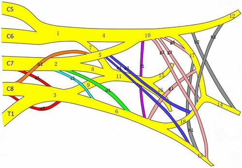
Results
Variations in the brachial plexus were identified in 40 of the 60 cadavers, yielding a prevalence rate of 66.7%. These variations were classified into root anomalies (2.1%), trunk anomalies (8.5%), division anomalies (2.1%), and cord anomalies (4.3%). Notably, anomalies in communicating branches were observed in 39 cadavers (83.0%): 14 with bilateral anomalies, 14 with anomalies on the left side, and 11 on the right side. These communicating branches formed connections between the roots and other segments, including trunks, cords, and terminal nerves, and involved the median, musculocutaneous, and ulnar nerves.
Conclusion
The frequency and diversity of brachial plexus variations, particularly in communicating branches, are significant in cadavers. It is imperative that these variations are carefully considered during the diagnostic process, treatment planning, and prior to procedures such as supraclavicular brachial plexus blocks and nerve transfers, to mitigate the risk of iatrogenic complications.
期刊介绍:
Microsurgery is an international and interdisciplinary publication of original contributions concerning surgery under microscopic magnification. Microsurgery publishes clinical studies, research papers, invited articles, relevant reviews, and other scholarly works from all related fields including orthopaedic surgery, otolaryngology, pediatric surgery, plastic surgery, urology, and vascular surgery.


 分享
分享
 求助内容:
求助内容: 应助结果提醒方式:
 扫码关注我们
