{"title":"患有膝关节骨性关节炎的女性在踝关节和膝关节肌肉结构以及足底压力分布方面的差异。","authors":"Nazli Busra Cigercioglu, Zilan Bazancir-Apaydin, Hakan Apaydin, Gul Baltaci, Hande Guney-Deniz","doi":"10.1002/jfa2.12028","DOIUrl":null,"url":null,"abstract":"<p><strong>Background: </strong>The aim of this study was to compare the plantar pressure distribution and knee and ankle muscle architecture in women with and without knee osteoarthritis (OA).</p><p><strong>Methods: </strong>Fifty women with knee OA (mean age = 52.11 ± 4.96 years, mean Body mass index (BMI) = 30.94 ± 4.23 kg/m<sup>2</sup>) and 50 healthy women as a control group (mean age = 50.93 ± 3.78 years, mean BMI = 29.06 ± 4.82 kg/m<sup>2</sup>) were included in the study. Ultrasonography was used to evaluate knee and ankle muscles architecture and femoral cartilage thickness. The plantar pressure distribution was evaluated using the Digital Biometry Scanning System and Milleri software (DIASU, Italy). Static foot posture was evaluated using the Foot Posture Index (FPI), and pain severity was assessed using the Visual Analog Scale.</p><p><strong>Results: </strong>The OA group exhibited lower muscle thickness in Rectus Femoris (RF) (p = 0.003), Vastus Medialis (VM) (p = 0.004), Vastus Lateralis (p = 0.023), and Peroneus Longus (p = 0.002), as well as lower Medial Gastrocnemius pennation angle (p = 0.049) and higher Fat thickness (FT) in RF (p = 0.033) and VM (p = 0.037) compared to the control group. The OA group showed thinner femoral cartilage thickness (p = 0.001) and higher pain severity (p = 0.001) than the control groups. FPI scores were higher (p = 0.001) in OA group compared to the control group. The plantar pressure distribution results indicated an increase in total surface (p = 0.027), total load (p = 0.002), medial load (p = 0.005), and lateral load (p = 0.002) on dominant side in OA group compared to the control group.</p><p><strong>Conclusions: </strong>Knee and ankle muscle architecture, knee extensor muscle FT, and plantar pressure distribution in the dominant foot differed in individuals with knee OA compared to the control group.</p>","PeriodicalId":49164,"journal":{"name":"Journal of Foot and Ankle Research","volume":"17 2","pages":"e12028"},"PeriodicalIF":2.2000,"publicationDate":"2024-06-01","publicationTypes":"Journal Article","fieldsOfStudy":null,"isOpenAccess":false,"openAccessPdf":"https://www.ncbi.nlm.nih.gov/pmc/articles/PMC11296719/pdf/","citationCount":"0","resultStr":"{\"title\":\"Differences in ankle and knee muscle architecture and plantar pressure distribution among women with knee osteoarthritis.\",\"authors\":\"Nazli Busra Cigercioglu, Zilan Bazancir-Apaydin, Hakan Apaydin, Gul Baltaci, Hande Guney-Deniz\",\"doi\":\"10.1002/jfa2.12028\",\"DOIUrl\":null,\"url\":null,\"abstract\":\"<p><strong>Background: </strong>The aim of this study was to compare the plantar pressure distribution and knee and ankle muscle architecture in women with and without knee osteoarthritis (OA).</p><p><strong>Methods: </strong>Fifty women with knee OA (mean age = 52.11 ± 4.96 years, mean Body mass index (BMI) = 30.94 ± 4.23 kg/m<sup>2</sup>) and 50 healthy women as a control group (mean age = 50.93 ± 3.78 years, mean BMI = 29.06 ± 4.82 kg/m<sup>2</sup>) were included in the study. Ultrasonography was used to evaluate knee and ankle muscles architecture and femoral cartilage thickness. The plantar pressure distribution was evaluated using the Digital Biometry Scanning System and Milleri software (DIASU, Italy). Static foot posture was evaluated using the Foot Posture Index (FPI), and pain severity was assessed using the Visual Analog Scale.</p><p><strong>Results: </strong>The OA group exhibited lower muscle thickness in Rectus Femoris (RF) (p = 0.003), Vastus Medialis (VM) (p = 0.004), Vastus Lateralis (p = 0.023), and Peroneus Longus (p = 0.002), as well as lower Medial Gastrocnemius pennation angle (p = 0.049) and higher Fat thickness (FT) in RF (p = 0.033) and VM (p = 0.037) compared to the control group. The OA group showed thinner femoral cartilage thickness (p = 0.001) and higher pain severity (p = 0.001) than the control groups. FPI scores were higher (p = 0.001) in OA group compared to the control group. The plantar pressure distribution results indicated an increase in total surface (p = 0.027), total load (p = 0.002), medial load (p = 0.005), and lateral load (p = 0.002) on dominant side in OA group compared to the control group.</p><p><strong>Conclusions: </strong>Knee and ankle muscle architecture, knee extensor muscle FT, and plantar pressure distribution in the dominant foot differed in individuals with knee OA compared to the control group.</p>\",\"PeriodicalId\":49164,\"journal\":{\"name\":\"Journal of Foot and Ankle Research\",\"volume\":\"17 2\",\"pages\":\"e12028\"},\"PeriodicalIF\":2.2000,\"publicationDate\":\"2024-06-01\",\"publicationTypes\":\"Journal Article\",\"fieldsOfStudy\":null,\"isOpenAccess\":false,\"openAccessPdf\":\"https://www.ncbi.nlm.nih.gov/pmc/articles/PMC11296719/pdf/\",\"citationCount\":\"0\",\"resultStr\":null,\"platform\":\"Semanticscholar\",\"paperid\":null,\"PeriodicalName\":\"Journal of Foot and Ankle Research\",\"FirstCategoryId\":\"3\",\"ListUrlMain\":\"https://doi.org/10.1002/jfa2.12028\",\"RegionNum\":3,\"RegionCategory\":\"医学\",\"ArticlePicture\":[],\"TitleCN\":null,\"AbstractTextCN\":null,\"PMCID\":null,\"EPubDate\":\"\",\"PubModel\":\"\",\"JCR\":\"Q1\",\"JCRName\":\"ORTHOPEDICS\",\"Score\":null,\"Total\":0}","platform":"Semanticscholar","paperid":null,"PeriodicalName":"Journal of Foot and Ankle Research","FirstCategoryId":"3","ListUrlMain":"https://doi.org/10.1002/jfa2.12028","RegionNum":3,"RegionCategory":"医学","ArticlePicture":[],"TitleCN":null,"AbstractTextCN":null,"PMCID":null,"EPubDate":"","PubModel":"","JCR":"Q1","JCRName":"ORTHOPEDICS","Score":null,"Total":0}
Differences in ankle and knee muscle architecture and plantar pressure distribution among women with knee osteoarthritis.
Background: The aim of this study was to compare the plantar pressure distribution and knee and ankle muscle architecture in women with and without knee osteoarthritis (OA).
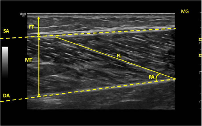
Methods: Fifty women with knee OA (mean age = 52.11 ± 4.96 years, mean Body mass index (BMI) = 30.94 ± 4.23 kg/m2) and 50 healthy women as a control group (mean age = 50.93 ± 3.78 years, mean BMI = 29.06 ± 4.82 kg/m2) were included in the study. Ultrasonography was used to evaluate knee and ankle muscles architecture and femoral cartilage thickness. The plantar pressure distribution was evaluated using the Digital Biometry Scanning System and Milleri software (DIASU, Italy). Static foot posture was evaluated using the Foot Posture Index (FPI), and pain severity was assessed using the Visual Analog Scale.
Results: The OA group exhibited lower muscle thickness in Rectus Femoris (RF) (p = 0.003), Vastus Medialis (VM) (p = 0.004), Vastus Lateralis (p = 0.023), and Peroneus Longus (p = 0.002), as well as lower Medial Gastrocnemius pennation angle (p = 0.049) and higher Fat thickness (FT) in RF (p = 0.033) and VM (p = 0.037) compared to the control group. The OA group showed thinner femoral cartilage thickness (p = 0.001) and higher pain severity (p = 0.001) than the control groups. FPI scores were higher (p = 0.001) in OA group compared to the control group. The plantar pressure distribution results indicated an increase in total surface (p = 0.027), total load (p = 0.002), medial load (p = 0.005), and lateral load (p = 0.002) on dominant side in OA group compared to the control group.
Conclusions: Knee and ankle muscle architecture, knee extensor muscle FT, and plantar pressure distribution in the dominant foot differed in individuals with knee OA compared to the control group.
期刊介绍:
Journal of Foot and Ankle Research, the official journal of the Australian Podiatry Association and The College of Podiatry (UK), is an open access journal that encompasses all aspects of policy, organisation, delivery and clinical practice related to the assessment, diagnosis, prevention and management of foot and ankle disorders.
Journal of Foot and Ankle Research covers a wide range of clinical subject areas, including diabetology, paediatrics, sports medicine, gerontology and geriatrics, foot surgery, physical therapy, dermatology, wound management, radiology, biomechanics and bioengineering, orthotics and prosthetics, as well the broad areas of epidemiology, policy, organisation and delivery of services related to foot and ankle care.
The journal encourages submissions from all health professionals who manage lower limb conditions, including podiatrists, nurses, physical therapists and physiotherapists, orthopaedists, manual therapists, medical specialists and general medical practitioners, as well as health service researchers concerned with foot and ankle care.
The Australian Podiatry Association and the College of Podiatry (UK) have reserve funds to cover the article-processing charge for manuscripts submitted by its members. Society members can email the appropriate contact at Australian Podiatry Association or The College of Podiatry to obtain the corresponding code to enter on submission.


 分享
分享
 求助内容:
求助内容: 应助结果提醒方式:
 扫码关注我们
