{"title":"人类颅脑拓扑的包容性解剖网络分析。","authors":"Tim Schuurman, Emiliano Bruner","doi":"10.1111/joa.14068","DOIUrl":null,"url":null,"abstract":"<p>The human brain's complex morphology is spatially constrained by numerous intrinsic and extrinsic physical interactions. Spatial constraints help to identify the source of morphological variability and can be investigated by employing anatomical network analysis. Here, a model of human craniocerebral topology is presented, based on the bony elements of the skull at birth and a previously designed model of the brain. The goal was to investigate the topological components fundamental to the craniocerebral geometric balance, to identify underlying phenotypic patterns of spatial arrangement, and to understand how these patterns might have influenced the evolution of human brain morphology. Analysis of the craniocerebral network model revealed that the combined structure of the body and lesser wings of the sphenoid bone, the parahippocampal gyrus, and the parietal and ethmoid bones are susceptible to sustain and apply major spatial constraints that are likely to limit or channel their morphological evolution. The results also showcase a high level of global integration and efficient diffusion of biomechanical forces across the craniocerebral system, a fundamental aspect of morphological variability in terms of plasticity. Finally, community detection in the craniocerebral system highlights the concurrence of a longitudinal and a vertical modular partition. The former reflects the distinct morphogenetic environments of the three endocranial fossae, while the latter corresponds to those of the basicranium and calvaria.</p>","PeriodicalId":14971,"journal":{"name":"Journal of Anatomy","volume":"245 5","pages":"686-698"},"PeriodicalIF":2.2000,"publicationDate":"2024-06-01","publicationTypes":"Journal Article","fieldsOfStudy":null,"isOpenAccess":false,"openAccessPdf":"","citationCount":"0","resultStr":"{\"title\":\"An inclusive anatomical network analysis of human craniocerebral topology\",\"authors\":\"Tim Schuurman, Emiliano Bruner\",\"doi\":\"10.1111/joa.14068\",\"DOIUrl\":null,\"url\":null,\"abstract\":\"<p>The human brain's complex morphology is spatially constrained by numerous intrinsic and extrinsic physical interactions. Spatial constraints help to identify the source of morphological variability and can be investigated by employing anatomical network analysis. Here, a model of human craniocerebral topology is presented, based on the bony elements of the skull at birth and a previously designed model of the brain. The goal was to investigate the topological components fundamental to the craniocerebral geometric balance, to identify underlying phenotypic patterns of spatial arrangement, and to understand how these patterns might have influenced the evolution of human brain morphology. Analysis of the craniocerebral network model revealed that the combined structure of the body and lesser wings of the sphenoid bone, the parahippocampal gyrus, and the parietal and ethmoid bones are susceptible to sustain and apply major spatial constraints that are likely to limit or channel their morphological evolution. The results also showcase a high level of global integration and efficient diffusion of biomechanical forces across the craniocerebral system, a fundamental aspect of morphological variability in terms of plasticity. Finally, community detection in the craniocerebral system highlights the concurrence of a longitudinal and a vertical modular partition. The former reflects the distinct morphogenetic environments of the three endocranial fossae, while the latter corresponds to those of the basicranium and calvaria.</p>\",\"PeriodicalId\":14971,\"journal\":{\"name\":\"Journal of Anatomy\",\"volume\":\"245 5\",\"pages\":\"686-698\"},\"PeriodicalIF\":2.2000,\"publicationDate\":\"2024-06-01\",\"publicationTypes\":\"Journal Article\",\"fieldsOfStudy\":null,\"isOpenAccess\":false,\"openAccessPdf\":\"\",\"citationCount\":\"0\",\"resultStr\":null,\"platform\":\"Semanticscholar\",\"paperid\":null,\"PeriodicalName\":\"Journal of Anatomy\",\"FirstCategoryId\":\"3\",\"ListUrlMain\":\"https://onlinelibrary.wiley.com/doi/10.1111/joa.14068\",\"RegionNum\":3,\"RegionCategory\":\"医学\",\"ArticlePicture\":[],\"TitleCN\":null,\"AbstractTextCN\":null,\"PMCID\":null,\"EPubDate\":\"\",\"PubModel\":\"\",\"JCR\":\"Q2\",\"JCRName\":\"ANATOMY & MORPHOLOGY\",\"Score\":null,\"Total\":0}","platform":"Semanticscholar","paperid":null,"PeriodicalName":"Journal of Anatomy","FirstCategoryId":"3","ListUrlMain":"https://onlinelibrary.wiley.com/doi/10.1111/joa.14068","RegionNum":3,"RegionCategory":"医学","ArticlePicture":[],"TitleCN":null,"AbstractTextCN":null,"PMCID":null,"EPubDate":"","PubModel":"","JCR":"Q2","JCRName":"ANATOMY & MORPHOLOGY","Score":null,"Total":0}
An inclusive anatomical network analysis of human craniocerebral topology
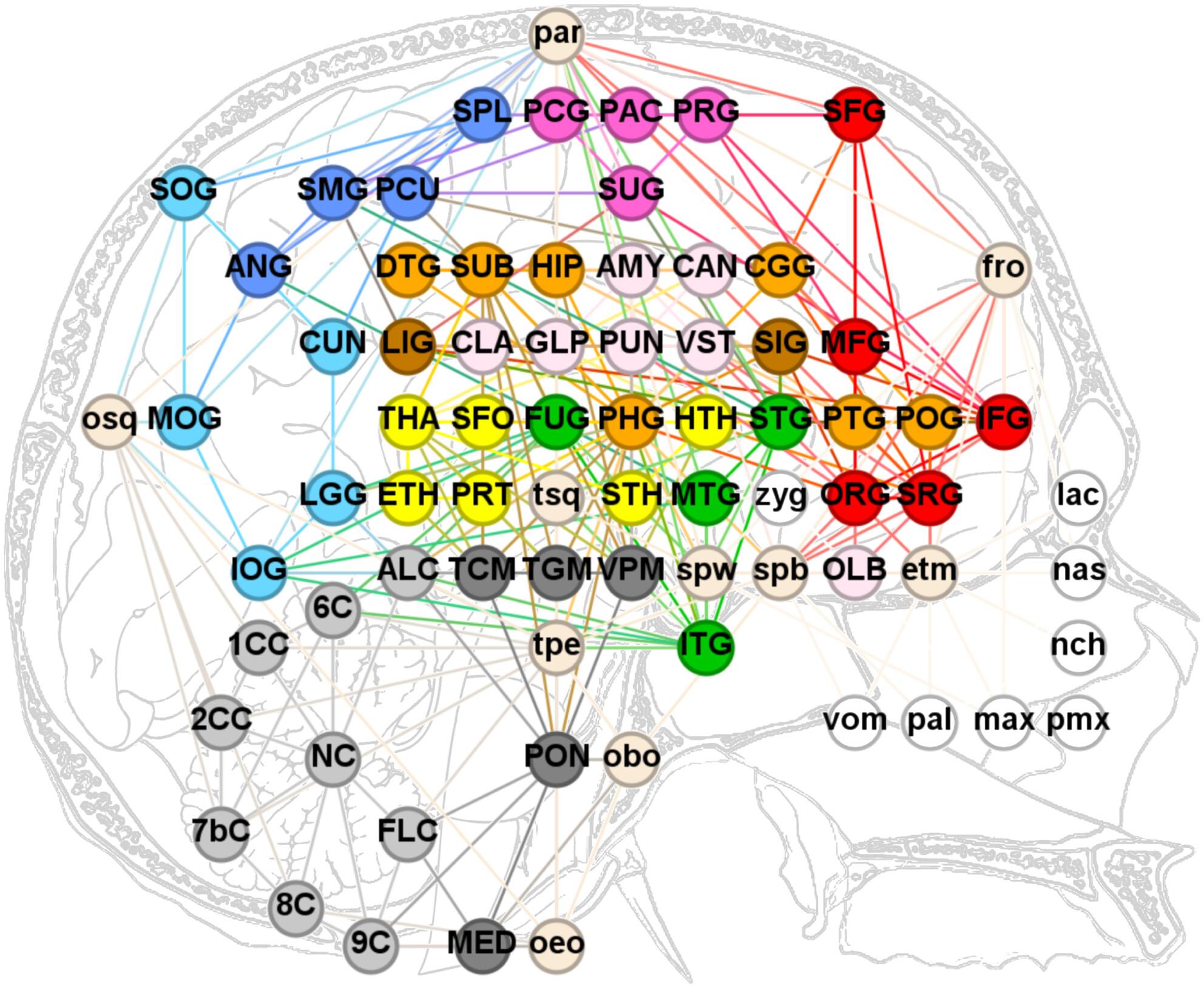
The human brain's complex morphology is spatially constrained by numerous intrinsic and extrinsic physical interactions. Spatial constraints help to identify the source of morphological variability and can be investigated by employing anatomical network analysis. Here, a model of human craniocerebral topology is presented, based on the bony elements of the skull at birth and a previously designed model of the brain. The goal was to investigate the topological components fundamental to the craniocerebral geometric balance, to identify underlying phenotypic patterns of spatial arrangement, and to understand how these patterns might have influenced the evolution of human brain morphology. Analysis of the craniocerebral network model revealed that the combined structure of the body and lesser wings of the sphenoid bone, the parahippocampal gyrus, and the parietal and ethmoid bones are susceptible to sustain and apply major spatial constraints that are likely to limit or channel their morphological evolution. The results also showcase a high level of global integration and efficient diffusion of biomechanical forces across the craniocerebral system, a fundamental aspect of morphological variability in terms of plasticity. Finally, community detection in the craniocerebral system highlights the concurrence of a longitudinal and a vertical modular partition. The former reflects the distinct morphogenetic environments of the three endocranial fossae, while the latter corresponds to those of the basicranium and calvaria.
期刊介绍:
Journal of Anatomy is an international peer-reviewed journal sponsored by the Anatomical Society. The journal publishes original papers, invited review articles and book reviews. Its main focus is to understand anatomy through an analysis of structure, function, development and evolution. Priority will be given to studies of that clearly articulate their relevance to the anatomical community. Focal areas include: experimental studies, contributions based on molecular and cell biology and on the application of modern imaging techniques and papers with novel methods or synthetic perspective on an anatomical system.
Studies that are essentially descriptive anatomy are appropriate only if they communicate clearly a broader functional or evolutionary significance. You must clearly state the broader implications of your work in the abstract.
We particularly welcome submissions in the following areas:
Cell biology and tissue architecture
Comparative functional morphology
Developmental biology
Evolutionary developmental biology
Evolutionary morphology
Functional human anatomy
Integrative vertebrate paleontology
Methodological innovations in anatomical research
Musculoskeletal system
Neuroanatomy and neurodegeneration
Significant advances in anatomical education.


 分享
分享
 求助内容:
求助内容: 应助结果提醒方式:
 扫码关注我们
