Eren Ogut, Serdar Bagci, Gonul Kotil Aslan, Pedram Turkoglu, Merve Falay, Cagatay Barut
{"title":"胸小肌双侧非典型起源的病例报告。","authors":"Eren Ogut, Serdar Bagci, Gonul Kotil Aslan, Pedram Turkoglu, Merve Falay, Cagatay Barut","doi":"10.1007/s00276-024-03407-9","DOIUrl":null,"url":null,"abstract":"<p><strong>Purpose: </strong>In the existing literature, various insertion variations and classifications for the Pectoralis Minor (PMi) muscle have been reported. However, there is limited information on inferior origin variations of the PMi muscles and a certain classification is lacking.</p><p><strong>Case presentation: </strong>During routine cadaver dissection of an adult male, variations in the origin of the bilateral PMi muscles were identified. Morphometric measurements of the PMi were conducted using ImageJ software, and the unusual origin patterns of the PMi were categorized into specific types. The PMi muscle demonstrated a bilateral variations. On the right side, the PMi displays a bifid structure comprising medial and lateral fibers. The left PMi originate from the superolateral margins of the 4th to 6th costae and terminate at the anterosuperior surface of the coracoid process. The length of the right medial fiber before merging was 5.67 ± 0.04 cm, while that of the right lateral fiber was 6.68 ± 0.05 cm. The distance between the two fibers was measured as 0.43 cm, with a length of 3.33 ± 0.02 cm. The length and diameter of the muscle fibers extending to the 6th costa were 2.63 ± 0.01 cm and 0.46 cm, respectively.</p><p><strong>Conclusions: </strong>Potential variations in PMi arising from impairment during development may occasionally manifest as asymptomatic conditions or predispose individuals to shoulder impingement, rotator cuff dysfunction, shoulder-related disorders, and functional impairments. Therefore, careful attention to this variation is considered in surgical planning.</p>","PeriodicalId":49461,"journal":{"name":"Surgical and Radiologic Anatomy","volume":" ","pages":"1373-1378"},"PeriodicalIF":1.2000,"publicationDate":"2024-08-01","publicationTypes":"Journal Article","fieldsOfStudy":null,"isOpenAccess":false,"openAccessPdf":"","citationCount":"0","resultStr":"{\"title\":\"A case report of bilateral atypical origin of pectoralis minor.\",\"authors\":\"Eren Ogut, Serdar Bagci, Gonul Kotil Aslan, Pedram Turkoglu, Merve Falay, Cagatay Barut\",\"doi\":\"10.1007/s00276-024-03407-9\",\"DOIUrl\":null,\"url\":null,\"abstract\":\"<p><strong>Purpose: </strong>In the existing literature, various insertion variations and classifications for the Pectoralis Minor (PMi) muscle have been reported. However, there is limited information on inferior origin variations of the PMi muscles and a certain classification is lacking.</p><p><strong>Case presentation: </strong>During routine cadaver dissection of an adult male, variations in the origin of the bilateral PMi muscles were identified. Morphometric measurements of the PMi were conducted using ImageJ software, and the unusual origin patterns of the PMi were categorized into specific types. The PMi muscle demonstrated a bilateral variations. On the right side, the PMi displays a bifid structure comprising medial and lateral fibers. The left PMi originate from the superolateral margins of the 4th to 6th costae and terminate at the anterosuperior surface of the coracoid process. The length of the right medial fiber before merging was 5.67 ± 0.04 cm, while that of the right lateral fiber was 6.68 ± 0.05 cm. The distance between the two fibers was measured as 0.43 cm, with a length of 3.33 ± 0.02 cm. The length and diameter of the muscle fibers extending to the 6th costa were 2.63 ± 0.01 cm and 0.46 cm, respectively.</p><p><strong>Conclusions: </strong>Potential variations in PMi arising from impairment during development may occasionally manifest as asymptomatic conditions or predispose individuals to shoulder impingement, rotator cuff dysfunction, shoulder-related disorders, and functional impairments. Therefore, careful attention to this variation is considered in surgical planning.</p>\",\"PeriodicalId\":49461,\"journal\":{\"name\":\"Surgical and Radiologic Anatomy\",\"volume\":\" \",\"pages\":\"1373-1378\"},\"PeriodicalIF\":1.2000,\"publicationDate\":\"2024-08-01\",\"publicationTypes\":\"Journal Article\",\"fieldsOfStudy\":null,\"isOpenAccess\":false,\"openAccessPdf\":\"\",\"citationCount\":\"0\",\"resultStr\":null,\"platform\":\"Semanticscholar\",\"paperid\":null,\"PeriodicalName\":\"Surgical and Radiologic Anatomy\",\"FirstCategoryId\":\"3\",\"ListUrlMain\":\"https://doi.org/10.1007/s00276-024-03407-9\",\"RegionNum\":4,\"RegionCategory\":\"医学\",\"ArticlePicture\":[],\"TitleCN\":null,\"AbstractTextCN\":null,\"PMCID\":null,\"EPubDate\":\"2024/6/10 0:00:00\",\"PubModel\":\"Epub\",\"JCR\":\"Q2\",\"JCRName\":\"Medicine\",\"Score\":null,\"Total\":0}","platform":"Semanticscholar","paperid":null,"PeriodicalName":"Surgical and Radiologic Anatomy","FirstCategoryId":"3","ListUrlMain":"https://doi.org/10.1007/s00276-024-03407-9","RegionNum":4,"RegionCategory":"医学","ArticlePicture":[],"TitleCN":null,"AbstractTextCN":null,"PMCID":null,"EPubDate":"2024/6/10 0:00:00","PubModel":"Epub","JCR":"Q2","JCRName":"Medicine","Score":null,"Total":0}
A case report of bilateral atypical origin of pectoralis minor.
Purpose: In the existing literature, various insertion variations and classifications for the Pectoralis Minor (PMi) muscle have been reported. However, there is limited information on inferior origin variations of the PMi muscles and a certain classification is lacking.
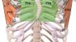
Case presentation: During routine cadaver dissection of an adult male, variations in the origin of the bilateral PMi muscles were identified. Morphometric measurements of the PMi were conducted using ImageJ software, and the unusual origin patterns of the PMi were categorized into specific types. The PMi muscle demonstrated a bilateral variations. On the right side, the PMi displays a bifid structure comprising medial and lateral fibers. The left PMi originate from the superolateral margins of the 4th to 6th costae and terminate at the anterosuperior surface of the coracoid process. The length of the right medial fiber before merging was 5.67 ± 0.04 cm, while that of the right lateral fiber was 6.68 ± 0.05 cm. The distance between the two fibers was measured as 0.43 cm, with a length of 3.33 ± 0.02 cm. The length and diameter of the muscle fibers extending to the 6th costa were 2.63 ± 0.01 cm and 0.46 cm, respectively.
Conclusions: Potential variations in PMi arising from impairment during development may occasionally manifest as asymptomatic conditions or predispose individuals to shoulder impingement, rotator cuff dysfunction, shoulder-related disorders, and functional impairments. Therefore, careful attention to this variation is considered in surgical planning.
期刊介绍:
Anatomy is a morphological science which cannot fail to interest the clinician. The practical application of anatomical research to clinical problems necessitates special adaptation and selectivity in choosing from numerous international works. Although there is a tendency to believe that meaningful advances in anatomy are unlikely, constant revision is necessary. Surgical and Radiologic Anatomy, the first international journal of Clinical anatomy has been created in this spirit.
Its goal is to serve clinicians, regardless of speciality-physicians, surgeons, radiologists or other specialists-as an indispensable aid with which they can improve their knowledge of anatomy. Each issue includes: Original papers, review articles, articles on the anatomical bases of medical, surgical and radiological techniques, articles of normal radiologic anatomy, brief reviews of anatomical publications of clinical interest.
Particular attention is given to high quality illustrations, which are indispensable for a better understanding of anatomical problems.
Surgical and Radiologic Anatomy is a journal written by anatomists for clinicians with a special interest in anatomy.


 分享
分享
 求助内容:
求助内容: 应助结果提醒方式:
 扫码关注我们
