{"title":"胃底-胃黏膜系的胃上皮肿瘤:低不典型性分化型胃肿瘤的代表,Ki67 可能有助于鉴别。","authors":"Houqiang Li, Lanqing Zheng, Guodong Zhong, Xunbin Yu, Xia Zhang, Linying Chen, Xin Chen","doi":"10.3389/pore.2024.1611734","DOIUrl":null,"url":null,"abstract":"<p><strong>Background: </strong>Gastric epithelial neoplasm of the fundic-gland mucosa lineages (GEN-FGMLs) are rare forms of gastric tumors that encompass oxyntic gland adenoma (OGA), gastric adenocarcinoma of the fundic-gland type (GA-FG), and gastric adenocarcinoma of the fundic-gland mucosa type (GA-FGM). There is no consensus on the cause, classification, and clinicopathological features of GEN-FGMLs, and misdiagnosis is common because of similarities in symptoms.</p><p><strong>Methods: </strong>37 cases diagnosed with GEN-FGMLs were included in this study. H&E-stained slides were reviewed and clinicopathological parameters were recorded. Immunohistochemical staining was conducted for MUC2, MUC5AC, MUC6, CD10, CD56, synaptophysin, chromograninA, p53, Ki67, pepsinogen-I, H<sup>+</sup>/K<sup>+</sup>-ATPase and Desmin.</p><p><strong>Results: </strong>The patients' ages ranged from 42 to 79 years, with a median age of 60. 17 were male and 20 were female. Morphologically, 19 OGAs, 16 GA-FGs, and two GA-FGMs were identified. Histopathological similarities exist between OGA, GA-FG, and GA-FGM. The tumors demonstrated well-formed glands, expanding with dense growth patterns comprising pale, blue-grey columnar cells with mild nuclear atypia. These cells resembled fundic gland cells. None of the OGA invaded the submucosal layer. The normal gastric pit epithelium covered the entire surface of the OGA and GA-FG, but the dysplasia pit epithelium covered the GA-FGM. Non-atrophic gastritis was observed in more than half of the background mucosa. All cases were diffusely positive for MUC6 and pepsinogen-I on immunohistochemistry. H<sup>+</sup>/K<sup>+</sup>-ATPase staining was negative or showed a scattered pattern in most cases. MUC5AC was expressed on the surface of GA-FGMs. p53 was focally expressed and the Ki67 index was low (1%-20%). Compared with OGA, GA-FG and GA-FGM were more prominent in the macroscopic view (<i>p</i> < 0.05) and had larger sizes (<i>p</i> < 0.0001). Additionally, GA-FG and GA-FGM exhibited higher Ki67 indices than OGA (<i>p</i> < 0.0001). Specimens with Ki-67 proliferation indices >2.5% and size >4.5 mm are more likely to be diagnosed with GA-FG and GA-FGM than OGA.</p><p><strong>Conclusion: </strong>GEN-FGMLs are group of well-differentiated gastric tumors with favourable biological behaviours, low cellular atypia, and low proliferation. Immunohistochemistry is critical for confirming diagnosis. Compared with OGA, GA-FG and GA-FGM have larger sizes and higher Ki67 proliferation indices, indicating that they play a critical role in the identification of GEN-FGML. Pathologists and endoscopists should be cautious to prevent misdiagnosis and overtreatment, especially in biopsy specimens.</p>","PeriodicalId":19981,"journal":{"name":"Pathology & Oncology Research","volume":"30 ","pages":"1611734"},"PeriodicalIF":2.7000,"publicationDate":"2024-05-30","publicationTypes":"Journal Article","fieldsOfStudy":null,"isOpenAccess":false,"openAccessPdf":"https://www.ncbi.nlm.nih.gov/pmc/articles/PMC11169639/pdf/","citationCount":"0","resultStr":"{\"title\":\"Gastric epithelial neoplasm of fundic-gland mucosa lineage: representative of the low atypia differentiated gastric tumor and Ki67 may help in their identification.\",\"authors\":\"Houqiang Li, Lanqing Zheng, Guodong Zhong, Xunbin Yu, Xia Zhang, Linying Chen, Xin Chen\",\"doi\":\"10.3389/pore.2024.1611734\",\"DOIUrl\":null,\"url\":null,\"abstract\":\"<p><strong>Background: </strong>Gastric epithelial neoplasm of the fundic-gland mucosa lineages (GEN-FGMLs) are rare forms of gastric tumors that encompass oxyntic gland adenoma (OGA), gastric adenocarcinoma of the fundic-gland type (GA-FG), and gastric adenocarcinoma of the fundic-gland mucosa type (GA-FGM). There is no consensus on the cause, classification, and clinicopathological features of GEN-FGMLs, and misdiagnosis is common because of similarities in symptoms.</p><p><strong>Methods: </strong>37 cases diagnosed with GEN-FGMLs were included in this study. H&E-stained slides were reviewed and clinicopathological parameters were recorded. Immunohistochemical staining was conducted for MUC2, MUC5AC, MUC6, CD10, CD56, synaptophysin, chromograninA, p53, Ki67, pepsinogen-I, H<sup>+</sup>/K<sup>+</sup>-ATPase and Desmin.</p><p><strong>Results: </strong>The patients' ages ranged from 42 to 79 years, with a median age of 60. 17 were male and 20 were female. Morphologically, 19 OGAs, 16 GA-FGs, and two GA-FGMs were identified. Histopathological similarities exist between OGA, GA-FG, and GA-FGM. The tumors demonstrated well-formed glands, expanding with dense growth patterns comprising pale, blue-grey columnar cells with mild nuclear atypia. These cells resembled fundic gland cells. None of the OGA invaded the submucosal layer. The normal gastric pit epithelium covered the entire surface of the OGA and GA-FG, but the dysplasia pit epithelium covered the GA-FGM. Non-atrophic gastritis was observed in more than half of the background mucosa. All cases were diffusely positive for MUC6 and pepsinogen-I on immunohistochemistry. H<sup>+</sup>/K<sup>+</sup>-ATPase staining was negative or showed a scattered pattern in most cases. MUC5AC was expressed on the surface of GA-FGMs. p53 was focally expressed and the Ki67 index was low (1%-20%). Compared with OGA, GA-FG and GA-FGM were more prominent in the macroscopic view (<i>p</i> < 0.05) and had larger sizes (<i>p</i> < 0.0001). Additionally, GA-FG and GA-FGM exhibited higher Ki67 indices than OGA (<i>p</i> < 0.0001). Specimens with Ki-67 proliferation indices >2.5% and size >4.5 mm are more likely to be diagnosed with GA-FG and GA-FGM than OGA.</p><p><strong>Conclusion: </strong>GEN-FGMLs are group of well-differentiated gastric tumors with favourable biological behaviours, low cellular atypia, and low proliferation. Immunohistochemistry is critical for confirming diagnosis. Compared with OGA, GA-FG and GA-FGM have larger sizes and higher Ki67 proliferation indices, indicating that they play a critical role in the identification of GEN-FGML. Pathologists and endoscopists should be cautious to prevent misdiagnosis and overtreatment, especially in biopsy specimens.</p>\",\"PeriodicalId\":19981,\"journal\":{\"name\":\"Pathology & Oncology Research\",\"volume\":\"30 \",\"pages\":\"1611734\"},\"PeriodicalIF\":2.7000,\"publicationDate\":\"2024-05-30\",\"publicationTypes\":\"Journal Article\",\"fieldsOfStudy\":null,\"isOpenAccess\":false,\"openAccessPdf\":\"https://www.ncbi.nlm.nih.gov/pmc/articles/PMC11169639/pdf/\",\"citationCount\":\"0\",\"resultStr\":null,\"platform\":\"Semanticscholar\",\"paperid\":null,\"PeriodicalName\":\"Pathology & Oncology Research\",\"FirstCategoryId\":\"3\",\"ListUrlMain\":\"https://doi.org/10.3389/pore.2024.1611734\",\"RegionNum\":4,\"RegionCategory\":\"医学\",\"ArticlePicture\":[],\"TitleCN\":null,\"AbstractTextCN\":null,\"PMCID\":null,\"EPubDate\":\"2024/1/1 0:00:00\",\"PubModel\":\"eCollection\",\"JCR\":\"Q3\",\"JCRName\":\"ONCOLOGY\",\"Score\":null,\"Total\":0}","platform":"Semanticscholar","paperid":null,"PeriodicalName":"Pathology & Oncology Research","FirstCategoryId":"3","ListUrlMain":"https://doi.org/10.3389/pore.2024.1611734","RegionNum":4,"RegionCategory":"医学","ArticlePicture":[],"TitleCN":null,"AbstractTextCN":null,"PMCID":null,"EPubDate":"2024/1/1 0:00:00","PubModel":"eCollection","JCR":"Q3","JCRName":"ONCOLOGY","Score":null,"Total":0}
Gastric epithelial neoplasm of fundic-gland mucosa lineage: representative of the low atypia differentiated gastric tumor and Ki67 may help in their identification.
Background: Gastric epithelial neoplasm of the fundic-gland mucosa lineages (GEN-FGMLs) are rare forms of gastric tumors that encompass oxyntic gland adenoma (OGA), gastric adenocarcinoma of the fundic-gland type (GA-FG), and gastric adenocarcinoma of the fundic-gland mucosa type (GA-FGM). There is no consensus on the cause, classification, and clinicopathological features of GEN-FGMLs, and misdiagnosis is common because of similarities in symptoms.
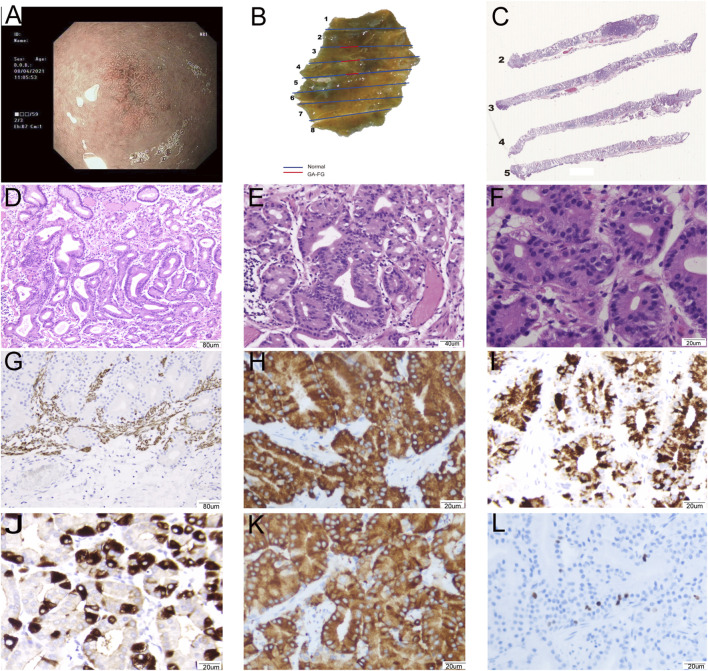
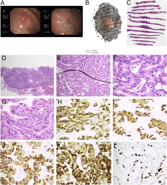
Methods: 37 cases diagnosed with GEN-FGMLs were included in this study. H&E-stained slides were reviewed and clinicopathological parameters were recorded. Immunohistochemical staining was conducted for MUC2, MUC5AC, MUC6, CD10, CD56, synaptophysin, chromograninA, p53, Ki67, pepsinogen-I, H+/K+-ATPase and Desmin.
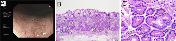
Results: The patients' ages ranged from 42 to 79 years, with a median age of 60. 17 were male and 20 were female. Morphologically, 19 OGAs, 16 GA-FGs, and two GA-FGMs were identified. Histopathological similarities exist between OGA, GA-FG, and GA-FGM. The tumors demonstrated well-formed glands, expanding with dense growth patterns comprising pale, blue-grey columnar cells with mild nuclear atypia. These cells resembled fundic gland cells. None of the OGA invaded the submucosal layer. The normal gastric pit epithelium covered the entire surface of the OGA and GA-FG, but the dysplasia pit epithelium covered the GA-FGM. Non-atrophic gastritis was observed in more than half of the background mucosa. All cases were diffusely positive for MUC6 and pepsinogen-I on immunohistochemistry. H+/K+-ATPase staining was negative or showed a scattered pattern in most cases. MUC5AC was expressed on the surface of GA-FGMs. p53 was focally expressed and the Ki67 index was low (1%-20%). Compared with OGA, GA-FG and GA-FGM were more prominent in the macroscopic view (p < 0.05) and had larger sizes (p < 0.0001). Additionally, GA-FG and GA-FGM exhibited higher Ki67 indices than OGA (p < 0.0001). Specimens with Ki-67 proliferation indices >2.5% and size >4.5 mm are more likely to be diagnosed with GA-FG and GA-FGM than OGA.
Conclusion: GEN-FGMLs are group of well-differentiated gastric tumors with favourable biological behaviours, low cellular atypia, and low proliferation. Immunohistochemistry is critical for confirming diagnosis. Compared with OGA, GA-FG and GA-FGM have larger sizes and higher Ki67 proliferation indices, indicating that they play a critical role in the identification of GEN-FGML. Pathologists and endoscopists should be cautious to prevent misdiagnosis and overtreatment, especially in biopsy specimens.
期刊介绍:
Pathology & Oncology Research (POR) is an interdisciplinary Journal at the interface of pathology and oncology including the preclinical and translational research, diagnostics and therapy. Furthermore, POR is an international forum for the rapid communication of reviews, original research, critical and topical reports with excellence and novelty. Published quarterly, POR is dedicated to keeping scientists informed of developments on the selected biomedical fields bridging the gap between basic research and clinical medicine. It is a special aim for POR to promote pathological and oncological publishing activity of colleagues in the Central and East European region. The journal will be of interest to pathologists, and a broad range of experimental and clinical oncologists, and related experts. POR is supported by an acknowledged international advisory board and the Arányi Fundation for modern pathology.


 分享
分享
 求助内容:
求助内容: 应助结果提醒方式:
 扫码关注我们
