Salvatore Marino, Guillaume Dannhoff, Christophe Destrieux, Igor Lima Maldonado
{"title":"脑岛的额叶经手术入路:从手术引导的解剖解剖中构建心理图像。","authors":"Salvatore Marino, Guillaume Dannhoff, Christophe Destrieux, Igor Lima Maldonado","doi":"10.1007/s00276-024-03409-7","DOIUrl":null,"url":null,"abstract":"<p><strong>Background: </strong>Performing transopercular frontal approaches to the insula, widely used in glioma surgeries, necessitates a meticulous understanding of both cortical and subcortical neuroanatomy. This precision is vital for preserving essential structures and accurately interpreting the results of direct electrical stimulation. Nevertheless, acquiring a compelling mental image of the anatomy of this region can be challenging due to several factors, among which stand out its complexity and the fact that white matter fasciculi are imperceptible to the naked eye in the living brain.</p><p><strong>Aim: </strong>In an effort to optimize the study of the anatomy relevant to this topic, we performed a procedure-guided laboratory study using subpial dissection, fiber dissection, vascular coloration, and stereoscopic photography in a \"real-life\" surgical perspective.</p><p><strong>Methods: </strong>Nine cerebral specimens obtained from body donation were extracted and fixed in formalin. Colored silicone injection and a variant of Klinglers's technique were used to demonstrate vascular and white matter structures, respectively. We dissected and photographed the specimens in a supero-antero-lateral view to reproduce the surgeon's viewpoint. The anatomy related to the development of the surgical corridor and resection cavity was documented using both standard photography and the red-cyan anaglyph technique.</p><p><strong>Results: </strong>The anatomy of frontal transopercular approaches to the insula involved elements of different natures-leptomeningeal, cortical, vascular, and fascicular-combining in the surgical field in a complex disposition. The disposition of these structures was successfully demonstrated through the aforementioned anatomical techniques. Among the main structures in or around the surgical corridor, the orbital, triangular, and opercular portions of the inferior frontal gyrus are critical landmarks in the cortical stage, as well as the leptomeninges of the Sylvian fissure and the M2-M4 branches of the middle cerebral artery in the subpial dissection stage, and the inferior fronto-occipital, uncinate and arcuate fasciculi, and the corona radiata in establishing the deep limits of resection.</p><p><strong>Conclusions: </strong>Procedure-guided study of cerebral hemispheres associating subpial, vascular, and fiber dissection from a surgical standpoint is a powerful tool for the realistic study of the surgical anatomy relevant to frontal transopercular approaches to the insula.</p>","PeriodicalId":49461,"journal":{"name":"Surgical and Radiologic Anatomy","volume":" ","pages":"1331-1344"},"PeriodicalIF":1.2000,"publicationDate":"2024-08-01","publicationTypes":"Journal Article","fieldsOfStudy":null,"isOpenAccess":false,"openAccessPdf":"","citationCount":"0","resultStr":"{\"title\":\"Frontal trans opercular approaches to the insula: building the mental picture from procedure-guided anatomical dissection.\",\"authors\":\"Salvatore Marino, Guillaume Dannhoff, Christophe Destrieux, Igor Lima Maldonado\",\"doi\":\"10.1007/s00276-024-03409-7\",\"DOIUrl\":null,\"url\":null,\"abstract\":\"<p><strong>Background: </strong>Performing transopercular frontal approaches to the insula, widely used in glioma surgeries, necessitates a meticulous understanding of both cortical and subcortical neuroanatomy. This precision is vital for preserving essential structures and accurately interpreting the results of direct electrical stimulation. Nevertheless, acquiring a compelling mental image of the anatomy of this region can be challenging due to several factors, among which stand out its complexity and the fact that white matter fasciculi are imperceptible to the naked eye in the living brain.</p><p><strong>Aim: </strong>In an effort to optimize the study of the anatomy relevant to this topic, we performed a procedure-guided laboratory study using subpial dissection, fiber dissection, vascular coloration, and stereoscopic photography in a \\\"real-life\\\" surgical perspective.</p><p><strong>Methods: </strong>Nine cerebral specimens obtained from body donation were extracted and fixed in formalin. Colored silicone injection and a variant of Klinglers's technique were used to demonstrate vascular and white matter structures, respectively. We dissected and photographed the specimens in a supero-antero-lateral view to reproduce the surgeon's viewpoint. The anatomy related to the development of the surgical corridor and resection cavity was documented using both standard photography and the red-cyan anaglyph technique.</p><p><strong>Results: </strong>The anatomy of frontal transopercular approaches to the insula involved elements of different natures-leptomeningeal, cortical, vascular, and fascicular-combining in the surgical field in a complex disposition. The disposition of these structures was successfully demonstrated through the aforementioned anatomical techniques. Among the main structures in or around the surgical corridor, the orbital, triangular, and opercular portions of the inferior frontal gyrus are critical landmarks in the cortical stage, as well as the leptomeninges of the Sylvian fissure and the M2-M4 branches of the middle cerebral artery in the subpial dissection stage, and the inferior fronto-occipital, uncinate and arcuate fasciculi, and the corona radiata in establishing the deep limits of resection.</p><p><strong>Conclusions: </strong>Procedure-guided study of cerebral hemispheres associating subpial, vascular, and fiber dissection from a surgical standpoint is a powerful tool for the realistic study of the surgical anatomy relevant to frontal transopercular approaches to the insula.</p>\",\"PeriodicalId\":49461,\"journal\":{\"name\":\"Surgical and Radiologic Anatomy\",\"volume\":\" \",\"pages\":\"1331-1344\"},\"PeriodicalIF\":1.2000,\"publicationDate\":\"2024-08-01\",\"publicationTypes\":\"Journal Article\",\"fieldsOfStudy\":null,\"isOpenAccess\":false,\"openAccessPdf\":\"\",\"citationCount\":\"0\",\"resultStr\":null,\"platform\":\"Semanticscholar\",\"paperid\":null,\"PeriodicalName\":\"Surgical and Radiologic Anatomy\",\"FirstCategoryId\":\"3\",\"ListUrlMain\":\"https://doi.org/10.1007/s00276-024-03409-7\",\"RegionNum\":4,\"RegionCategory\":\"医学\",\"ArticlePicture\":[],\"TitleCN\":null,\"AbstractTextCN\":null,\"PMCID\":null,\"EPubDate\":\"2024/6/13 0:00:00\",\"PubModel\":\"Epub\",\"JCR\":\"Q2\",\"JCRName\":\"Medicine\",\"Score\":null,\"Total\":0}","platform":"Semanticscholar","paperid":null,"PeriodicalName":"Surgical and Radiologic Anatomy","FirstCategoryId":"3","ListUrlMain":"https://doi.org/10.1007/s00276-024-03409-7","RegionNum":4,"RegionCategory":"医学","ArticlePicture":[],"TitleCN":null,"AbstractTextCN":null,"PMCID":null,"EPubDate":"2024/6/13 0:00:00","PubModel":"Epub","JCR":"Q2","JCRName":"Medicine","Score":null,"Total":0}
Frontal trans opercular approaches to the insula: building the mental picture from procedure-guided anatomical dissection.
Background: Performing transopercular frontal approaches to the insula, widely used in glioma surgeries, necessitates a meticulous understanding of both cortical and subcortical neuroanatomy. This precision is vital for preserving essential structures and accurately interpreting the results of direct electrical stimulation. Nevertheless, acquiring a compelling mental image of the anatomy of this region can be challenging due to several factors, among which stand out its complexity and the fact that white matter fasciculi are imperceptible to the naked eye in the living brain.
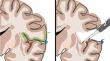
Aim: In an effort to optimize the study of the anatomy relevant to this topic, we performed a procedure-guided laboratory study using subpial dissection, fiber dissection, vascular coloration, and stereoscopic photography in a "real-life" surgical perspective.
Methods: Nine cerebral specimens obtained from body donation were extracted and fixed in formalin. Colored silicone injection and a variant of Klinglers's technique were used to demonstrate vascular and white matter structures, respectively. We dissected and photographed the specimens in a supero-antero-lateral view to reproduce the surgeon's viewpoint. The anatomy related to the development of the surgical corridor and resection cavity was documented using both standard photography and the red-cyan anaglyph technique.
Results: The anatomy of frontal transopercular approaches to the insula involved elements of different natures-leptomeningeal, cortical, vascular, and fascicular-combining in the surgical field in a complex disposition. The disposition of these structures was successfully demonstrated through the aforementioned anatomical techniques. Among the main structures in or around the surgical corridor, the orbital, triangular, and opercular portions of the inferior frontal gyrus are critical landmarks in the cortical stage, as well as the leptomeninges of the Sylvian fissure and the M2-M4 branches of the middle cerebral artery in the subpial dissection stage, and the inferior fronto-occipital, uncinate and arcuate fasciculi, and the corona radiata in establishing the deep limits of resection.
Conclusions: Procedure-guided study of cerebral hemispheres associating subpial, vascular, and fiber dissection from a surgical standpoint is a powerful tool for the realistic study of the surgical anatomy relevant to frontal transopercular approaches to the insula.
期刊介绍:
Anatomy is a morphological science which cannot fail to interest the clinician. The practical application of anatomical research to clinical problems necessitates special adaptation and selectivity in choosing from numerous international works. Although there is a tendency to believe that meaningful advances in anatomy are unlikely, constant revision is necessary. Surgical and Radiologic Anatomy, the first international journal of Clinical anatomy has been created in this spirit.
Its goal is to serve clinicians, regardless of speciality-physicians, surgeons, radiologists or other specialists-as an indispensable aid with which they can improve their knowledge of anatomy. Each issue includes: Original papers, review articles, articles on the anatomical bases of medical, surgical and radiological techniques, articles of normal radiologic anatomy, brief reviews of anatomical publications of clinical interest.
Particular attention is given to high quality illustrations, which are indispensable for a better understanding of anatomical problems.
Surgical and Radiologic Anatomy is a journal written by anatomists for clinicians with a special interest in anatomy.


 分享
分享
 求助内容:
求助内容: 应助结果提醒方式:
 扫码关注我们
