P Jeanneton, A De Barros, S Alshehri, V Poulet, Z Cavallier, A Prevost, R Lopez
{"title":"下颌骨和软组织的动脉血管化。解剖研究。","authors":"P Jeanneton, A De Barros, S Alshehri, V Poulet, Z Cavallier, A Prevost, R Lopez","doi":"10.1007/s00276-024-03320-1","DOIUrl":null,"url":null,"abstract":"<p><strong>Purpose: </strong>The literature has for too long described the arterial supply of the mandible as coming from a single artery, the inferior alveolar artery, and being of the terminal type. Rather, it appears to come from an extensive and complex arterial network dependent on the lingual, facial, and maxillary arteries and their collateral branches. Our study aims to confirm and demonstrate the arterial vascular richness of the mandible and to establish arterial mapping.</p><p><strong>Methods: </strong>The arterial vascularization of the mandible was revealed in six anatomic specimens after performing selective injections of the lingual, facial, and maxillary arteries with different dyes. A specimen was injected intra-arterially with colored latex at the level of the maxillary artery for a morphometric study.</p><p><strong>Results: </strong>Eighteen selective arterial injections were performed on six anatomic specimens. The mucocutaneous, musculoperiosteal, and intramedullary vascularizations were analyzed. Each of the arteries has a defined and delimited cutaneo-mucous vascular territory. The facial and maxillary arteries supply the musculoperiosteal vascularization of the mandible from the condyle to the symphysis. The lingual artery supplies only the inner cortex of the parasymphyseal and symphyseal regions. The facial and maxillary arteries provide intramedullary vascularization from the angle of the mandible to the parasymphysis. The vascularization of the symphysis depends on the facial artery. No staining was found in the condyle region. Neoprene latex injection was performed on an anatomic specimen, revealing a permeable anastomosis between the inferior alveolar and facial arteries.</p><p><strong>Conclusion: </strong>The arterial vascularization of the mandible is dependent on the maxillary, facial, and lingual arteries. This is a network vasculature. This study makes it possible to establish an arterial map of the mandible. The presence of an anastomosis between the inferior alveolar artery and the facial artery confirms the existence of dynamic and borrowed vascularization. Knowledge of this arterial system makes it possible to adapt maxillofacial surgical care and to anticipate possible intraoperative complications.</p>","PeriodicalId":49461,"journal":{"name":"Surgical and Radiologic Anatomy","volume":" ","pages":"1219-1230"},"PeriodicalIF":1.2000,"publicationDate":"2024-08-01","publicationTypes":"Journal Article","fieldsOfStudy":null,"isOpenAccess":false,"openAccessPdf":"","citationCount":"0","resultStr":"{\"title\":\"Arterial vascularization of the mandible and soft tissues. Anatomic study.\",\"authors\":\"P Jeanneton, A De Barros, S Alshehri, V Poulet, Z Cavallier, A Prevost, R Lopez\",\"doi\":\"10.1007/s00276-024-03320-1\",\"DOIUrl\":null,\"url\":null,\"abstract\":\"<p><strong>Purpose: </strong>The literature has for too long described the arterial supply of the mandible as coming from a single artery, the inferior alveolar artery, and being of the terminal type. Rather, it appears to come from an extensive and complex arterial network dependent on the lingual, facial, and maxillary arteries and their collateral branches. Our study aims to confirm and demonstrate the arterial vascular richness of the mandible and to establish arterial mapping.</p><p><strong>Methods: </strong>The arterial vascularization of the mandible was revealed in six anatomic specimens after performing selective injections of the lingual, facial, and maxillary arteries with different dyes. A specimen was injected intra-arterially with colored latex at the level of the maxillary artery for a morphometric study.</p><p><strong>Results: </strong>Eighteen selective arterial injections were performed on six anatomic specimens. The mucocutaneous, musculoperiosteal, and intramedullary vascularizations were analyzed. Each of the arteries has a defined and delimited cutaneo-mucous vascular territory. The facial and maxillary arteries supply the musculoperiosteal vascularization of the mandible from the condyle to the symphysis. The lingual artery supplies only the inner cortex of the parasymphyseal and symphyseal regions. The facial and maxillary arteries provide intramedullary vascularization from the angle of the mandible to the parasymphysis. The vascularization of the symphysis depends on the facial artery. No staining was found in the condyle region. Neoprene latex injection was performed on an anatomic specimen, revealing a permeable anastomosis between the inferior alveolar and facial arteries.</p><p><strong>Conclusion: </strong>The arterial vascularization of the mandible is dependent on the maxillary, facial, and lingual arteries. This is a network vasculature. This study makes it possible to establish an arterial map of the mandible. The presence of an anastomosis between the inferior alveolar artery and the facial artery confirms the existence of dynamic and borrowed vascularization. Knowledge of this arterial system makes it possible to adapt maxillofacial surgical care and to anticipate possible intraoperative complications.</p>\",\"PeriodicalId\":49461,\"journal\":{\"name\":\"Surgical and Radiologic Anatomy\",\"volume\":\" \",\"pages\":\"1219-1230\"},\"PeriodicalIF\":1.2000,\"publicationDate\":\"2024-08-01\",\"publicationTypes\":\"Journal Article\",\"fieldsOfStudy\":null,\"isOpenAccess\":false,\"openAccessPdf\":\"\",\"citationCount\":\"0\",\"resultStr\":null,\"platform\":\"Semanticscholar\",\"paperid\":null,\"PeriodicalName\":\"Surgical and Radiologic Anatomy\",\"FirstCategoryId\":\"3\",\"ListUrlMain\":\"https://doi.org/10.1007/s00276-024-03320-1\",\"RegionNum\":4,\"RegionCategory\":\"医学\",\"ArticlePicture\":[],\"TitleCN\":null,\"AbstractTextCN\":null,\"PMCID\":null,\"EPubDate\":\"2024/6/18 0:00:00\",\"PubModel\":\"Epub\",\"JCR\":\"Q2\",\"JCRName\":\"Medicine\",\"Score\":null,\"Total\":0}","platform":"Semanticscholar","paperid":null,"PeriodicalName":"Surgical and Radiologic Anatomy","FirstCategoryId":"3","ListUrlMain":"https://doi.org/10.1007/s00276-024-03320-1","RegionNum":4,"RegionCategory":"医学","ArticlePicture":[],"TitleCN":null,"AbstractTextCN":null,"PMCID":null,"EPubDate":"2024/6/18 0:00:00","PubModel":"Epub","JCR":"Q2","JCRName":"Medicine","Score":null,"Total":0}
Arterial vascularization of the mandible and soft tissues. Anatomic study.
Purpose: The literature has for too long described the arterial supply of the mandible as coming from a single artery, the inferior alveolar artery, and being of the terminal type. Rather, it appears to come from an extensive and complex arterial network dependent on the lingual, facial, and maxillary arteries and their collateral branches. Our study aims to confirm and demonstrate the arterial vascular richness of the mandible and to establish arterial mapping.
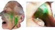
Methods: The arterial vascularization of the mandible was revealed in six anatomic specimens after performing selective injections of the lingual, facial, and maxillary arteries with different dyes. A specimen was injected intra-arterially with colored latex at the level of the maxillary artery for a morphometric study.
Results: Eighteen selective arterial injections were performed on six anatomic specimens. The mucocutaneous, musculoperiosteal, and intramedullary vascularizations were analyzed. Each of the arteries has a defined and delimited cutaneo-mucous vascular territory. The facial and maxillary arteries supply the musculoperiosteal vascularization of the mandible from the condyle to the symphysis. The lingual artery supplies only the inner cortex of the parasymphyseal and symphyseal regions. The facial and maxillary arteries provide intramedullary vascularization from the angle of the mandible to the parasymphysis. The vascularization of the symphysis depends on the facial artery. No staining was found in the condyle region. Neoprene latex injection was performed on an anatomic specimen, revealing a permeable anastomosis between the inferior alveolar and facial arteries.
Conclusion: The arterial vascularization of the mandible is dependent on the maxillary, facial, and lingual arteries. This is a network vasculature. This study makes it possible to establish an arterial map of the mandible. The presence of an anastomosis between the inferior alveolar artery and the facial artery confirms the existence of dynamic and borrowed vascularization. Knowledge of this arterial system makes it possible to adapt maxillofacial surgical care and to anticipate possible intraoperative complications.
期刊介绍:
Anatomy is a morphological science which cannot fail to interest the clinician. The practical application of anatomical research to clinical problems necessitates special adaptation and selectivity in choosing from numerous international works. Although there is a tendency to believe that meaningful advances in anatomy are unlikely, constant revision is necessary. Surgical and Radiologic Anatomy, the first international journal of Clinical anatomy has been created in this spirit.
Its goal is to serve clinicians, regardless of speciality-physicians, surgeons, radiologists or other specialists-as an indispensable aid with which they can improve their knowledge of anatomy. Each issue includes: Original papers, review articles, articles on the anatomical bases of medical, surgical and radiological techniques, articles of normal radiologic anatomy, brief reviews of anatomical publications of clinical interest.
Particular attention is given to high quality illustrations, which are indispensable for a better understanding of anatomical problems.
Surgical and Radiologic Anatomy is a journal written by anatomists for clinicians with a special interest in anatomy.


 分享
分享
 求助内容:
求助内容: 应助结果提醒方式:
 扫码关注我们
