{"title":"基于双能 CT 和先进的多参数 MRI 的胰腺纤维化成像生物标记物在慢性胰腺炎严重程度分级中的实用性。","authors":"Mohak Narang, Anup Singh, Soumya Jagannath Mahapatra, Deepak Gunjan, Sanjay Sharma, Deep Narayan Srivastava, Rajni Yadav, Nihar Ranjan Dash, Virinder Kumar Bansal, Ravindra Mohan Pandey, Pramod Kumar Garg, Kumble Seetharama Madhusudhan","doi":"10.1007/s00261-024-04443-0","DOIUrl":null,"url":null,"abstract":"<div><h3>Purpose</h3><p>To non-invasively quantify pancreatic fibrosis and grade severity of chronic pancreatitis (CP) on dual-energy CT (DECT) and multiparametric MRI (mpMRI).</p><h3>Methods</h3><p>We included 72 patients (mean age:30years; 59 men) with suspected or confirmed CP from December 2019 to December 2021 graded as equivocal(<i>n</i> = 20), mild(<i>n</i> = 18), and moderate-marked(<i>n</i> = 34) using composite imaging and endoscopic ultrasound criteria. Study patients underwent multiphasic DECT and mpMRI of the abdomen. Normalized iodine concentration(NIC) and fat fraction(FF) on 6-minute delayed DECT, and T1 relaxation time(T1Rt), extracellular volume fraction(ECVf), intravoxel incoherent motion-based perfusion fraction(PF), and magnetization transfer ratio(MTR) on mpMRI of pancreas were compared. 20 renal donors(for DECT) and 20 patients with renal mass(for mpMRI) served as controls.</p><h3>Results</h3><p>NIC of pancreas in controls and progressive grades of CP were 0.24 ± 0.05, 0.80 ± 0.18, 1.06 ± 0.23, 1.40 ± 0.36, FF were 9.28 <i>±</i> 5.89, 14.19 <i>±</i> 5.29, 17.31 <i>±</i> 5.99, 29.32 <i>±</i> 12.22, T1Rt were 590.11 ± 61.13, 801.93 ± 211.01, 1006.79 ± 352.18, 1388.01 ± 312.23ms, ECVf were 0.07 ± 0.03, 0.30 ± 0.12, 0.41 ± 0.12, 0.53 ± 0.13, PF were 0.38 ± 0.04, 0.28 ± 0.07, 0.25 ± 0.09, 0.21 ± 0.05 and MTR were 0.12 ± 0.03, 0.15 ± 0.06, 0.21 ± 0.07, 0.26 ± 0.06, respectively. There were significant differences for all quantitative parameters between controls and mild CP; for NIC, PF, and ECVf between controls and progressive CP grades (<i>p</i> < 0.05). Area under curve for NIC, FF, T1Rt, ECVf, PF, and MTR in differentiating controls and mild CP were 1.00, 0.86, 0.95, 1.00, 0.90 and 0.84 respectively and for NIC, FF, ECVf and PF in differentiating controls and equivocal CP were 1.00, 0.76, 0.95 and 0.92 respectively.</p><h3>Conclusion</h3><p>DECT and mpMRI were useful in quantifying pancreatic fibrosis and grading the severity of CP. NIC was the most accurate marker.</p></div>","PeriodicalId":7126,"journal":{"name":"Abdominal Radiology","volume":"49 10","pages":"3528 - 3539"},"PeriodicalIF":2.2000,"publicationDate":"2024-06-20","publicationTypes":"Journal Article","fieldsOfStudy":null,"isOpenAccess":false,"openAccessPdf":"","citationCount":"0","resultStr":"{\"title\":\"Utility of dual-energy CT and advanced multiparametric MRI based imaging biomarkers of pancreatic fibrosis in grading the severity of chronic pancreatitis\",\"authors\":\"Mohak Narang, Anup Singh, Soumya Jagannath Mahapatra, Deepak Gunjan, Sanjay Sharma, Deep Narayan Srivastava, Rajni Yadav, Nihar Ranjan Dash, Virinder Kumar Bansal, Ravindra Mohan Pandey, Pramod Kumar Garg, Kumble Seetharama Madhusudhan\",\"doi\":\"10.1007/s00261-024-04443-0\",\"DOIUrl\":null,\"url\":null,\"abstract\":\"<div><h3>Purpose</h3><p>To non-invasively quantify pancreatic fibrosis and grade severity of chronic pancreatitis (CP) on dual-energy CT (DECT) and multiparametric MRI (mpMRI).</p><h3>Methods</h3><p>We included 72 patients (mean age:30years; 59 men) with suspected or confirmed CP from December 2019 to December 2021 graded as equivocal(<i>n</i> = 20), mild(<i>n</i> = 18), and moderate-marked(<i>n</i> = 34) using composite imaging and endoscopic ultrasound criteria. Study patients underwent multiphasic DECT and mpMRI of the abdomen. Normalized iodine concentration(NIC) and fat fraction(FF) on 6-minute delayed DECT, and T1 relaxation time(T1Rt), extracellular volume fraction(ECVf), intravoxel incoherent motion-based perfusion fraction(PF), and magnetization transfer ratio(MTR) on mpMRI of pancreas were compared. 20 renal donors(for DECT) and 20 patients with renal mass(for mpMRI) served as controls.</p><h3>Results</h3><p>NIC of pancreas in controls and progressive grades of CP were 0.24 ± 0.05, 0.80 ± 0.18, 1.06 ± 0.23, 1.40 ± 0.36, FF were 9.28 <i>±</i> 5.89, 14.19 <i>±</i> 5.29, 17.31 <i>±</i> 5.99, 29.32 <i>±</i> 12.22, T1Rt were 590.11 ± 61.13, 801.93 ± 211.01, 1006.79 ± 352.18, 1388.01 ± 312.23ms, ECVf were 0.07 ± 0.03, 0.30 ± 0.12, 0.41 ± 0.12, 0.53 ± 0.13, PF were 0.38 ± 0.04, 0.28 ± 0.07, 0.25 ± 0.09, 0.21 ± 0.05 and MTR were 0.12 ± 0.03, 0.15 ± 0.06, 0.21 ± 0.07, 0.26 ± 0.06, respectively. There were significant differences for all quantitative parameters between controls and mild CP; for NIC, PF, and ECVf between controls and progressive CP grades (<i>p</i> < 0.05). Area under curve for NIC, FF, T1Rt, ECVf, PF, and MTR in differentiating controls and mild CP were 1.00, 0.86, 0.95, 1.00, 0.90 and 0.84 respectively and for NIC, FF, ECVf and PF in differentiating controls and equivocal CP were 1.00, 0.76, 0.95 and 0.92 respectively.</p><h3>Conclusion</h3><p>DECT and mpMRI were useful in quantifying pancreatic fibrosis and grading the severity of CP. NIC was the most accurate marker.</p></div>\",\"PeriodicalId\":7126,\"journal\":{\"name\":\"Abdominal Radiology\",\"volume\":\"49 10\",\"pages\":\"3528 - 3539\"},\"PeriodicalIF\":2.2000,\"publicationDate\":\"2024-06-20\",\"publicationTypes\":\"Journal Article\",\"fieldsOfStudy\":null,\"isOpenAccess\":false,\"openAccessPdf\":\"\",\"citationCount\":\"0\",\"resultStr\":null,\"platform\":\"Semanticscholar\",\"paperid\":null,\"PeriodicalName\":\"Abdominal Radiology\",\"FirstCategoryId\":\"3\",\"ListUrlMain\":\"https://link.springer.com/article/10.1007/s00261-024-04443-0\",\"RegionNum\":3,\"RegionCategory\":\"医学\",\"ArticlePicture\":[],\"TitleCN\":null,\"AbstractTextCN\":null,\"PMCID\":null,\"EPubDate\":\"\",\"PubModel\":\"\",\"JCR\":\"Q2\",\"JCRName\":\"RADIOLOGY, NUCLEAR MEDICINE & MEDICAL IMAGING\",\"Score\":null,\"Total\":0}","platform":"Semanticscholar","paperid":null,"PeriodicalName":"Abdominal Radiology","FirstCategoryId":"3","ListUrlMain":"https://link.springer.com/article/10.1007/s00261-024-04443-0","RegionNum":3,"RegionCategory":"医学","ArticlePicture":[],"TitleCN":null,"AbstractTextCN":null,"PMCID":null,"EPubDate":"","PubModel":"","JCR":"Q2","JCRName":"RADIOLOGY, NUCLEAR MEDICINE & MEDICAL IMAGING","Score":null,"Total":0}
Utility of dual-energy CT and advanced multiparametric MRI based imaging biomarkers of pancreatic fibrosis in grading the severity of chronic pancreatitis
Purpose
To non-invasively quantify pancreatic fibrosis and grade severity of chronic pancreatitis (CP) on dual-energy CT (DECT) and multiparametric MRI (mpMRI).
Methods
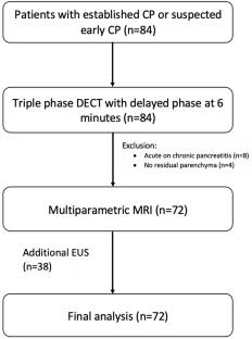
We included 72 patients (mean age:30years; 59 men) with suspected or confirmed CP from December 2019 to December 2021 graded as equivocal(n = 20), mild(n = 18), and moderate-marked(n = 34) using composite imaging and endoscopic ultrasound criteria. Study patients underwent multiphasic DECT and mpMRI of the abdomen. Normalized iodine concentration(NIC) and fat fraction(FF) on 6-minute delayed DECT, and T1 relaxation time(T1Rt), extracellular volume fraction(ECVf), intravoxel incoherent motion-based perfusion fraction(PF), and magnetization transfer ratio(MTR) on mpMRI of pancreas were compared. 20 renal donors(for DECT) and 20 patients with renal mass(for mpMRI) served as controls.
Results
NIC of pancreas in controls and progressive grades of CP were 0.24 ± 0.05, 0.80 ± 0.18, 1.06 ± 0.23, 1.40 ± 0.36, FF were 9.28 ± 5.89, 14.19 ± 5.29, 17.31 ± 5.99, 29.32 ± 12.22, T1Rt were 590.11 ± 61.13, 801.93 ± 211.01, 1006.79 ± 352.18, 1388.01 ± 312.23ms, ECVf were 0.07 ± 0.03, 0.30 ± 0.12, 0.41 ± 0.12, 0.53 ± 0.13, PF were 0.38 ± 0.04, 0.28 ± 0.07, 0.25 ± 0.09, 0.21 ± 0.05 and MTR were 0.12 ± 0.03, 0.15 ± 0.06, 0.21 ± 0.07, 0.26 ± 0.06, respectively. There were significant differences for all quantitative parameters between controls and mild CP; for NIC, PF, and ECVf between controls and progressive CP grades (p < 0.05). Area under curve for NIC, FF, T1Rt, ECVf, PF, and MTR in differentiating controls and mild CP were 1.00, 0.86, 0.95, 1.00, 0.90 and 0.84 respectively and for NIC, FF, ECVf and PF in differentiating controls and equivocal CP were 1.00, 0.76, 0.95 and 0.92 respectively.
Conclusion
DECT and mpMRI were useful in quantifying pancreatic fibrosis and grading the severity of CP. NIC was the most accurate marker.
期刊介绍:
Abdominal Radiology seeks to meet the professional needs of the abdominal radiologist by publishing clinically pertinent original, review and practice related articles on the gastrointestinal and genitourinary tracts and abdominal interventional and radiologic procedures. Case reports are generally not accepted unless they are the first report of a new disease or condition, or part of a special solicited section.
Reasons to Publish Your Article in Abdominal Radiology:
· Official journal of the Society of Abdominal Radiology (SAR)
· Published in Cooperation with:
European Society of Gastrointestinal and Abdominal Radiology (ESGAR)
European Society of Urogenital Radiology (ESUR)
Asian Society of Abdominal Radiology (ASAR)
· Efficient handling and Expeditious review
· Author feedback is provided in a mentoring style
· Global readership
· Readers can earn CME credits


 分享
分享
 求助内容:
求助内容: 应助结果提醒方式:
 扫码关注我们
