{"title":"扫描电子显微镜中石棉纤维的透射电子成像和衍射。","authors":"Jason D. Holm and Elisabeth Mansfield","doi":"10.1039/D4AY00555D","DOIUrl":null,"url":null,"abstract":"<p >Test protocols for airborne clearance of asbestos abatement sites define the collection, imaging and quantification of asbestos with transmission electron microscopy (TEM). Since those protocols were developed 35 years ago, scanning electron microscope (SEM) capabilities have significantly improved and expanded, with improvements in image spatial resolution, elemental analysis, and transmission electron diffraction capabilities. This contribution demonstrates transmission electron imaging and diffraction using NIST Asbestos Standard Reference Materials and a conventional SEM to provide comparable identification and quantification capabilities in the SEM as the current regulatory methods based on TEM techniques. In particular, we demonstrate that the 0.53 nm layer line spacing that is characteristic of asbestos can be quantified using different detection methods, and that other identifying diffraction signatures of chrysotile are readily obtained. The results demonstrate a viable alternative to the current TEM-based methods for asbestos identification and classification.</p>","PeriodicalId":64,"journal":{"name":"Analytical Methods","volume":" 27","pages":" 4570-4581"},"PeriodicalIF":2.6000,"publicationDate":"2024-06-24","publicationTypes":"Journal Article","fieldsOfStudy":null,"isOpenAccess":false,"openAccessPdf":"","citationCount":"0","resultStr":"{\"title\":\"Transmission electron imaging and diffraction of asbestos fibers in a scanning electron microscope†\",\"authors\":\"Jason D. Holm and Elisabeth Mansfield\",\"doi\":\"10.1039/D4AY00555D\",\"DOIUrl\":null,\"url\":null,\"abstract\":\"<p >Test protocols for airborne clearance of asbestos abatement sites define the collection, imaging and quantification of asbestos with transmission electron microscopy (TEM). Since those protocols were developed 35 years ago, scanning electron microscope (SEM) capabilities have significantly improved and expanded, with improvements in image spatial resolution, elemental analysis, and transmission electron diffraction capabilities. This contribution demonstrates transmission electron imaging and diffraction using NIST Asbestos Standard Reference Materials and a conventional SEM to provide comparable identification and quantification capabilities in the SEM as the current regulatory methods based on TEM techniques. In particular, we demonstrate that the 0.53 nm layer line spacing that is characteristic of asbestos can be quantified using different detection methods, and that other identifying diffraction signatures of chrysotile are readily obtained. The results demonstrate a viable alternative to the current TEM-based methods for asbestos identification and classification.</p>\",\"PeriodicalId\":64,\"journal\":{\"name\":\"Analytical Methods\",\"volume\":\" 27\",\"pages\":\" 4570-4581\"},\"PeriodicalIF\":2.6000,\"publicationDate\":\"2024-06-24\",\"publicationTypes\":\"Journal Article\",\"fieldsOfStudy\":null,\"isOpenAccess\":false,\"openAccessPdf\":\"\",\"citationCount\":\"0\",\"resultStr\":null,\"platform\":\"Semanticscholar\",\"paperid\":null,\"PeriodicalName\":\"Analytical Methods\",\"FirstCategoryId\":\"92\",\"ListUrlMain\":\"https://pubs.rsc.org/en/content/articlelanding/2024/ay/d4ay00555d\",\"RegionNum\":3,\"RegionCategory\":\"化学\",\"ArticlePicture\":[],\"TitleCN\":null,\"AbstractTextCN\":null,\"PMCID\":null,\"EPubDate\":\"\",\"PubModel\":\"\",\"JCR\":\"Q2\",\"JCRName\":\"CHEMISTRY, ANALYTICAL\",\"Score\":null,\"Total\":0}","platform":"Semanticscholar","paperid":null,"PeriodicalName":"Analytical Methods","FirstCategoryId":"92","ListUrlMain":"https://pubs.rsc.org/en/content/articlelanding/2024/ay/d4ay00555d","RegionNum":3,"RegionCategory":"化学","ArticlePicture":[],"TitleCN":null,"AbstractTextCN":null,"PMCID":null,"EPubDate":"","PubModel":"","JCR":"Q2","JCRName":"CHEMISTRY, ANALYTICAL","Score":null,"Total":0}
引用次数: 0
摘要
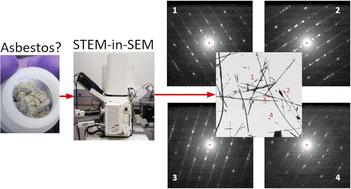
石棉清除现场的空气传播测试协议规定了利用透射电子显微镜(TEM)对石棉进行收集、成像和量化。自 35 年前制定这些规程以来,扫描电子显微镜(SEM)的功能得到了显著的改进和扩展,在图像空间分辨率、元素分析和透射电子衍射功能方面都有所提高。本文利用 NIST 石棉标准参考材料和传统扫描电子显微镜演示了透射电子成像和衍射,使扫描电子显微镜具有与目前基于 TEM 技术的监管方法相当的鉴定和量化能力。特别是,我们证明了可以使用不同的检测方法对石棉特有的 0.53 nm 层间距进行量化,并且可以轻松获得温石棉的其他识别衍射特征。这些结果表明,除了目前基于 TEM 的石棉鉴定和分类方法之外,还有一种可行的替代方法。
Transmission electron imaging and diffraction of asbestos fibers in a scanning electron microscope†
Test protocols for airborne clearance of asbestos abatement sites define the collection, imaging and quantification of asbestos with transmission electron microscopy (TEM). Since those protocols were developed 35 years ago, scanning electron microscope (SEM) capabilities have significantly improved and expanded, with improvements in image spatial resolution, elemental analysis, and transmission electron diffraction capabilities. This contribution demonstrates transmission electron imaging and diffraction using NIST Asbestos Standard Reference Materials and a conventional SEM to provide comparable identification and quantification capabilities in the SEM as the current regulatory methods based on TEM techniques. In particular, we demonstrate that the 0.53 nm layer line spacing that is characteristic of asbestos can be quantified using different detection methods, and that other identifying diffraction signatures of chrysotile are readily obtained. The results demonstrate a viable alternative to the current TEM-based methods for asbestos identification and classification.


 分享
分享
 求助内容:
求助内容: 应助结果提醒方式:
 扫码关注我们
