{"title":"COVID-19 睡眠障碍患者灰质的持续改变:一项为期 3 个月的纵向研究。","authors":"Kaixuan Zhou, Gaoxiong Duan, Ying Liu, Bei Peng, Xiaoyan Zhou, Lixia Qin, Lingyan Liang, Yichen Wei, Qingping Zhang, Xiaocheng Li, Haixia Qin, Yinqi Lai, Yian Lu, Yan Zhang, Jiazhu Huang, Jinli Huang, Yinfei Ouyang, Bolin Bin, Mingming Zhao, Jun Liu, Jianrong Yang, Demao Deng","doi":"10.4103/NRR.NRR-D-23-01651","DOIUrl":null,"url":null,"abstract":"<p><p>JOURNAL/nrgr/04.03/01300535-202510000-00030/figure1/v/2024-11-26T163120Z/r/image-tiff Sleep disturbances are among the most prevalent neuropsychiatric symptoms in individuals who have recovered from severe acute respiratory syndrome coronavirus 2 infections. Previous studies have demonstrated abnormal brain structures in patients with sleep disturbances who have recovered from coronavirus disease 2019 (COVID-19). However, neuroimaging studies on sleep disturbances caused by COVID-19 are scarce, and existing studies have primarily focused on the long-term effects of the virus, with minimal acute phase data. As a result, little is known about the pathophysiology of sleep disturbances in the acute phase of COVID-19. To address this issue, we designed a longitudinal study to investigate whether alterations in brain structure occur during the acute phase of infection, and verified the results using 3-month follow-up data. A total of 26 COVID-19 patients with sleep disturbances (aged 51.5 ± 13.57 years, 8 women and 18 men), 27 COVID-19 patients without sleep disturbances (aged 47.33 ± 15.98 years, 9 women and 18 men), and 31 age- and gender-matched healthy controls (aged 49.19 ± 17.51 years, 9 women and 22 men) were included in this study. Eleven COVID-19 patients with sleep disturbances were included in a longitudinal analysis. We found that COVID-19 patients with sleep disturbances exhibited brain structural changes in almost all brain lobes. The cortical thicknesses of the left pars opercularis and left precuneus were significantly negatively correlated with Pittsburgh Sleep Quality Index scores. Additionally, we observed changes in the volume of the hippocampus and its subfield regions in COVID-19 patients compared with the healthy controls. The 3-month follow-up data revealed indices of altered cerebral structure (cortical thickness, cortical grey matter volume, and cortical surface area) in the frontal-parietal cortex compared with the baseline in COVID-19 patients with sleep disturbances. Our findings indicate that the sleep disturbances patients had altered morphology in the cortical and hippocampal structures during the acute phase of infection and persistent changes in cortical regions at 3 months post-infection. These data improve our understanding of the pathophysiology of sleep disturbances caused by COVID-19.</p>","PeriodicalId":19113,"journal":{"name":"Neural Regeneration Research","volume":" ","pages":"3013-3024"},"PeriodicalIF":8.5000,"publicationDate":"2025-10-01","publicationTypes":"Journal Article","fieldsOfStudy":null,"isOpenAccess":false,"openAccessPdf":"https://www.ncbi.nlm.nih.gov/pmc/articles/PMC11826451/pdf/","citationCount":"0","resultStr":"{\"title\":\"Persistent alterations in gray matter in COVID-19 patients experiencing sleep disturbances: a 3-month longitudinal study.\",\"authors\":\"Kaixuan Zhou, Gaoxiong Duan, Ying Liu, Bei Peng, Xiaoyan Zhou, Lixia Qin, Lingyan Liang, Yichen Wei, Qingping Zhang, Xiaocheng Li, Haixia Qin, Yinqi Lai, Yian Lu, Yan Zhang, Jiazhu Huang, Jinli Huang, Yinfei Ouyang, Bolin Bin, Mingming Zhao, Jun Liu, Jianrong Yang, Demao Deng\",\"doi\":\"10.4103/NRR.NRR-D-23-01651\",\"DOIUrl\":null,\"url\":null,\"abstract\":\"<p><p>JOURNAL/nrgr/04.03/01300535-202510000-00030/figure1/v/2024-11-26T163120Z/r/image-tiff Sleep disturbances are among the most prevalent neuropsychiatric symptoms in individuals who have recovered from severe acute respiratory syndrome coronavirus 2 infections. Previous studies have demonstrated abnormal brain structures in patients with sleep disturbances who have recovered from coronavirus disease 2019 (COVID-19). However, neuroimaging studies on sleep disturbances caused by COVID-19 are scarce, and existing studies have primarily focused on the long-term effects of the virus, with minimal acute phase data. As a result, little is known about the pathophysiology of sleep disturbances in the acute phase of COVID-19. To address this issue, we designed a longitudinal study to investigate whether alterations in brain structure occur during the acute phase of infection, and verified the results using 3-month follow-up data. A total of 26 COVID-19 patients with sleep disturbances (aged 51.5 ± 13.57 years, 8 women and 18 men), 27 COVID-19 patients without sleep disturbances (aged 47.33 ± 15.98 years, 9 women and 18 men), and 31 age- and gender-matched healthy controls (aged 49.19 ± 17.51 years, 9 women and 22 men) were included in this study. Eleven COVID-19 patients with sleep disturbances were included in a longitudinal analysis. We found that COVID-19 patients with sleep disturbances exhibited brain structural changes in almost all brain lobes. The cortical thicknesses of the left pars opercularis and left precuneus were significantly negatively correlated with Pittsburgh Sleep Quality Index scores. Additionally, we observed changes in the volume of the hippocampus and its subfield regions in COVID-19 patients compared with the healthy controls. The 3-month follow-up data revealed indices of altered cerebral structure (cortical thickness, cortical grey matter volume, and cortical surface area) in the frontal-parietal cortex compared with the baseline in COVID-19 patients with sleep disturbances. Our findings indicate that the sleep disturbances patients had altered morphology in the cortical and hippocampal structures during the acute phase of infection and persistent changes in cortical regions at 3 months post-infection. These data improve our understanding of the pathophysiology of sleep disturbances caused by COVID-19.</p>\",\"PeriodicalId\":19113,\"journal\":{\"name\":\"Neural Regeneration Research\",\"volume\":\" \",\"pages\":\"3013-3024\"},\"PeriodicalIF\":8.5000,\"publicationDate\":\"2025-10-01\",\"publicationTypes\":\"Journal Article\",\"fieldsOfStudy\":null,\"isOpenAccess\":false,\"openAccessPdf\":\"https://www.ncbi.nlm.nih.gov/pmc/articles/PMC11826451/pdf/\",\"citationCount\":\"0\",\"resultStr\":null,\"platform\":\"Semanticscholar\",\"paperid\":null,\"PeriodicalName\":\"Neural Regeneration Research\",\"FirstCategoryId\":\"3\",\"ListUrlMain\":\"https://doi.org/10.4103/NRR.NRR-D-23-01651\",\"RegionNum\":2,\"RegionCategory\":\"医学\",\"ArticlePicture\":[],\"TitleCN\":null,\"AbstractTextCN\":null,\"PMCID\":null,\"EPubDate\":\"2024/6/26 0:00:00\",\"PubModel\":\"Epub\",\"JCR\":\"Q2\",\"JCRName\":\"CELL BIOLOGY\",\"Score\":null,\"Total\":0}","platform":"Semanticscholar","paperid":null,"PeriodicalName":"Neural Regeneration Research","FirstCategoryId":"3","ListUrlMain":"https://doi.org/10.4103/NRR.NRR-D-23-01651","RegionNum":2,"RegionCategory":"医学","ArticlePicture":[],"TitleCN":null,"AbstractTextCN":null,"PMCID":null,"EPubDate":"2024/6/26 0:00:00","PubModel":"Epub","JCR":"Q2","JCRName":"CELL BIOLOGY","Score":null,"Total":0}
Persistent alterations in gray matter in COVID-19 patients experiencing sleep disturbances: a 3-month longitudinal study.
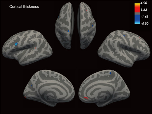
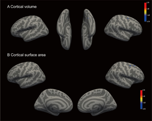
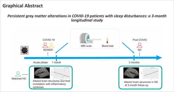
JOURNAL/nrgr/04.03/01300535-202510000-00030/figure1/v/2024-11-26T163120Z/r/image-tiff Sleep disturbances are among the most prevalent neuropsychiatric symptoms in individuals who have recovered from severe acute respiratory syndrome coronavirus 2 infections. Previous studies have demonstrated abnormal brain structures in patients with sleep disturbances who have recovered from coronavirus disease 2019 (COVID-19). However, neuroimaging studies on sleep disturbances caused by COVID-19 are scarce, and existing studies have primarily focused on the long-term effects of the virus, with minimal acute phase data. As a result, little is known about the pathophysiology of sleep disturbances in the acute phase of COVID-19. To address this issue, we designed a longitudinal study to investigate whether alterations in brain structure occur during the acute phase of infection, and verified the results using 3-month follow-up data. A total of 26 COVID-19 patients with sleep disturbances (aged 51.5 ± 13.57 years, 8 women and 18 men), 27 COVID-19 patients without sleep disturbances (aged 47.33 ± 15.98 years, 9 women and 18 men), and 31 age- and gender-matched healthy controls (aged 49.19 ± 17.51 years, 9 women and 22 men) were included in this study. Eleven COVID-19 patients with sleep disturbances were included in a longitudinal analysis. We found that COVID-19 patients with sleep disturbances exhibited brain structural changes in almost all brain lobes. The cortical thicknesses of the left pars opercularis and left precuneus were significantly negatively correlated with Pittsburgh Sleep Quality Index scores. Additionally, we observed changes in the volume of the hippocampus and its subfield regions in COVID-19 patients compared with the healthy controls. The 3-month follow-up data revealed indices of altered cerebral structure (cortical thickness, cortical grey matter volume, and cortical surface area) in the frontal-parietal cortex compared with the baseline in COVID-19 patients with sleep disturbances. Our findings indicate that the sleep disturbances patients had altered morphology in the cortical and hippocampal structures during the acute phase of infection and persistent changes in cortical regions at 3 months post-infection. These data improve our understanding of the pathophysiology of sleep disturbances caused by COVID-19.
期刊介绍:
Neural Regeneration Research (NRR) is the Open Access journal specializing in neural regeneration and indexed by SCI-E and PubMed. The journal is committed to publishing articles on basic pathobiology of injury, repair and protection to the nervous system, while considering preclinical and clinical trials targeted at improving traumatically injuried patients and patients with neurodegenerative diseases.


 分享
分享
 求助内容:
求助内容: 应助结果提醒方式:
 扫码关注我们
