Alastair David Bezzina, Jeremy Spiteri Bailey, Isaac Bertuello
{"title":"继发于疟疾的 II 型急性黄斑神经视网膜病变。","authors":"Alastair David Bezzina, Jeremy Spiteri Bailey, Isaac Bertuello","doi":"10.1155/2024/1577127","DOIUrl":null,"url":null,"abstract":"<p><p>To the best of our knowledge, we present the first case of type II acute macular neuroretinopathy (AMN) exhibiting in a patient suffering from malarial retinopathy concomitant with cerebral malaria acquired after travelling to West Africa without taking the necessary antimalarial prophylaxis. The patient complained of bilateral blurring of vision after being removed off sedation whilst at the intensive care unit. Subsequent examination revealed bilateral retinal haemorrhages, cotton-wool spots, and foveal pigmentary changes in keeping malarial retinopathy. Macular optical coherence tomography (OCT) revealed patchy hyperreflective changes at the level of the outer plexiform and outer nuclear layers (ONL) in keeping with the areas of deep capillary plexus flow void noted on OCT-angiography (OCT-A). This case report sheds more light on the extent of neurosensory retinal ischaemia in malarial retinopathy and showcases a new imaging biomarker which may be utilized in assessing and quantifying the functional deficit created by this disease.</p>","PeriodicalId":9603,"journal":{"name":"Case Reports in Ophthalmological Medicine","volume":"2024 ","pages":"1577127"},"PeriodicalIF":0.4000,"publicationDate":"2024-05-29","publicationTypes":"Journal Article","fieldsOfStudy":null,"isOpenAccess":false,"openAccessPdf":"https://www.ncbi.nlm.nih.gov/pmc/articles/PMC11208812/pdf/","citationCount":"0","resultStr":"{\"title\":\"Type II Acute Macular Neuroretinopathy Secondary to Malaria.\",\"authors\":\"Alastair David Bezzina, Jeremy Spiteri Bailey, Isaac Bertuello\",\"doi\":\"10.1155/2024/1577127\",\"DOIUrl\":null,\"url\":null,\"abstract\":\"<p><p>To the best of our knowledge, we present the first case of type II acute macular neuroretinopathy (AMN) exhibiting in a patient suffering from malarial retinopathy concomitant with cerebral malaria acquired after travelling to West Africa without taking the necessary antimalarial prophylaxis. The patient complained of bilateral blurring of vision after being removed off sedation whilst at the intensive care unit. Subsequent examination revealed bilateral retinal haemorrhages, cotton-wool spots, and foveal pigmentary changes in keeping malarial retinopathy. Macular optical coherence tomography (OCT) revealed patchy hyperreflective changes at the level of the outer plexiform and outer nuclear layers (ONL) in keeping with the areas of deep capillary plexus flow void noted on OCT-angiography (OCT-A). This case report sheds more light on the extent of neurosensory retinal ischaemia in malarial retinopathy and showcases a new imaging biomarker which may be utilized in assessing and quantifying the functional deficit created by this disease.</p>\",\"PeriodicalId\":9603,\"journal\":{\"name\":\"Case Reports in Ophthalmological Medicine\",\"volume\":\"2024 \",\"pages\":\"1577127\"},\"PeriodicalIF\":0.4000,\"publicationDate\":\"2024-05-29\",\"publicationTypes\":\"Journal Article\",\"fieldsOfStudy\":null,\"isOpenAccess\":false,\"openAccessPdf\":\"https://www.ncbi.nlm.nih.gov/pmc/articles/PMC11208812/pdf/\",\"citationCount\":\"0\",\"resultStr\":null,\"platform\":\"Semanticscholar\",\"paperid\":null,\"PeriodicalName\":\"Case Reports in Ophthalmological Medicine\",\"FirstCategoryId\":\"1085\",\"ListUrlMain\":\"https://doi.org/10.1155/2024/1577127\",\"RegionNum\":0,\"RegionCategory\":null,\"ArticlePicture\":[],\"TitleCN\":null,\"AbstractTextCN\":null,\"PMCID\":null,\"EPubDate\":\"2024/1/1 0:00:00\",\"PubModel\":\"eCollection\",\"JCR\":\"Q4\",\"JCRName\":\"OPHTHALMOLOGY\",\"Score\":null,\"Total\":0}","platform":"Semanticscholar","paperid":null,"PeriodicalName":"Case Reports in Ophthalmological Medicine","FirstCategoryId":"1085","ListUrlMain":"https://doi.org/10.1155/2024/1577127","RegionNum":0,"RegionCategory":null,"ArticlePicture":[],"TitleCN":null,"AbstractTextCN":null,"PMCID":null,"EPubDate":"2024/1/1 0:00:00","PubModel":"eCollection","JCR":"Q4","JCRName":"OPHTHALMOLOGY","Score":null,"Total":0}
引用次数: 0
摘要
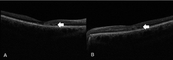
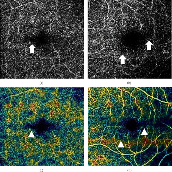
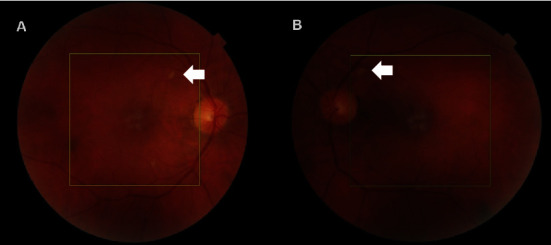
据我们所知,这是我们发现的首例Ⅱ型急性黄斑神经视网膜病变(AMN)病例,患者患有疟原虫性视网膜病变,同时伴有脑疟疾,在前往西非旅行时未采取必要的抗疟预防措施。患者在重症监护室解除镇静后,主诉双侧视力模糊。随后的检查发现双侧视网膜出血、棉絮状斑点和眼窝色素改变,表明患者患有疟原虫视网膜病变。黄斑光学相干断层扫描(OCT)显示,丛状外层和核外层(ONL)出现斑片状高反射变化,与 OCT 血管造影(OCT-A)显示的深层毛细血管丛流动空洞区域一致。本病例报告进一步揭示了疟原虫视网膜病变中神经感觉视网膜缺血的程度,并展示了一种新的成像生物标志物,可用于评估和量化该疾病造成的功能障碍。
Type II Acute Macular Neuroretinopathy Secondary to Malaria.
To the best of our knowledge, we present the first case of type II acute macular neuroretinopathy (AMN) exhibiting in a patient suffering from malarial retinopathy concomitant with cerebral malaria acquired after travelling to West Africa without taking the necessary antimalarial prophylaxis. The patient complained of bilateral blurring of vision after being removed off sedation whilst at the intensive care unit. Subsequent examination revealed bilateral retinal haemorrhages, cotton-wool spots, and foveal pigmentary changes in keeping malarial retinopathy. Macular optical coherence tomography (OCT) revealed patchy hyperreflective changes at the level of the outer plexiform and outer nuclear layers (ONL) in keeping with the areas of deep capillary plexus flow void noted on OCT-angiography (OCT-A). This case report sheds more light on the extent of neurosensory retinal ischaemia in malarial retinopathy and showcases a new imaging biomarker which may be utilized in assessing and quantifying the functional deficit created by this disease.


 分享
分享
 求助内容:
求助内容: 应助结果提醒方式:
 扫码关注我们
