Theresa J van Lith, Wouter M Sluis, Naomi T Wijers, Frederick J A Meijer, Karin Kamphuis-van Ulzen, Jeroen de Bresser, Jan Willem Dankbaar, Quirijn de Mast, Frederikus A Klok, Suzanne C Cannegieter, Marieke J H Wermer, Menno V Huisman, Anil M Tuladhar, H Bart van der Worp, Frank-Erik de Leeuw
{"title":"COVID-19住院患者脑血管磁共振成像标记物的流行率和3个月随访:CORONIS研究。","authors":"Theresa J van Lith, Wouter M Sluis, Naomi T Wijers, Frederick J A Meijer, Karin Kamphuis-van Ulzen, Jeroen de Bresser, Jan Willem Dankbaar, Quirijn de Mast, Frederikus A Klok, Suzanne C Cannegieter, Marieke J H Wermer, Menno V Huisman, Anil M Tuladhar, H Bart van der Worp, Frank-Erik de Leeuw","doi":"10.1007/s00234-024-03411-1","DOIUrl":null,"url":null,"abstract":"<p><strong>Purpose: </strong>To investigate the prevalence of cerebrovascular MRI markers in unselected patients hospitalized for COVID-19 (Coronavirus disease 2019), we compared these with healthy controls without previous SARS-CoV-2 infection or hospitalization and subsequently, investigated longitudinal (incidental) lesions in patients after three months.</p><p><strong>Methods: </strong>CORONIS (CORONavirus and Ischemic Stroke) was an observational cohort study in adult hospitalized patients for COVID-19 and controls without COVID-19, conducted between April 2021 and September 2022. Brain MRI was performed shortly after discharge and after 3 months. Outcomes included recent ischemic (DWI-positive) lesions, previous infarction, microbleeds, white matter hyperintensities (WMH) and intracerebral hemorrhage and were analysed with logistic regression to adjust for confounders.</p><p><strong>Results: </strong>125 patients with COVID-19 and 47 controls underwent brain MRI a median of 41.5 days after symptom onset. DWI-positive lesions were found in one patient (1%) and in one (2%) control, both clinically silent. WMH were more prevalent in patients (78%) than in controls (62%) (adjusted OR: 2.95 [95% CI: 1.07-8.57]), other cerebrovascular MRI markers did not differ. Prevalence of markers in ICU vs. non-ICU patients was similar. After three months, five patients (5%) had new cerebrovascular lesions, including DWI-positive lesions (1 patient, 1.0%), cerebral infarction (2 patients, 2.0%) and microbleeds (3 patients, 3.1%).</p><p><strong>Conclusion: </strong>Overall, we found no higher prevalence of cerebrovascular markers in unselected hospitalized COVID-19 patients compared to controls. The few incident DWI-lesions were most likely to be explained by risk-factors of small vessel disease. In the general hospitalized COVID-19 population, COVID-19 shows limited impact on cerebrovascular MRI markers shortly after hospitalization.</p>","PeriodicalId":19422,"journal":{"name":"Neuroradiology","volume":null,"pages":null},"PeriodicalIF":2.4000,"publicationDate":"2024-09-01","publicationTypes":"Journal Article","fieldsOfStudy":null,"isOpenAccess":false,"openAccessPdf":"https://www.ncbi.nlm.nih.gov/pmc/articles/PMC11322373/pdf/","citationCount":"0","resultStr":"{\"title\":\"Prevalence and 3-month follow-up of cerebrovascular MRI markers in hospitalized COVID-19 patients: the CORONIS study.\",\"authors\":\"Theresa J van Lith, Wouter M Sluis, Naomi T Wijers, Frederick J A Meijer, Karin Kamphuis-van Ulzen, Jeroen de Bresser, Jan Willem Dankbaar, Quirijn de Mast, Frederikus A Klok, Suzanne C Cannegieter, Marieke J H Wermer, Menno V Huisman, Anil M Tuladhar, H Bart van der Worp, Frank-Erik de Leeuw\",\"doi\":\"10.1007/s00234-024-03411-1\",\"DOIUrl\":null,\"url\":null,\"abstract\":\"<p><strong>Purpose: </strong>To investigate the prevalence of cerebrovascular MRI markers in unselected patients hospitalized for COVID-19 (Coronavirus disease 2019), we compared these with healthy controls without previous SARS-CoV-2 infection or hospitalization and subsequently, investigated longitudinal (incidental) lesions in patients after three months.</p><p><strong>Methods: </strong>CORONIS (CORONavirus and Ischemic Stroke) was an observational cohort study in adult hospitalized patients for COVID-19 and controls without COVID-19, conducted between April 2021 and September 2022. Brain MRI was performed shortly after discharge and after 3 months. Outcomes included recent ischemic (DWI-positive) lesions, previous infarction, microbleeds, white matter hyperintensities (WMH) and intracerebral hemorrhage and were analysed with logistic regression to adjust for confounders.</p><p><strong>Results: </strong>125 patients with COVID-19 and 47 controls underwent brain MRI a median of 41.5 days after symptom onset. DWI-positive lesions were found in one patient (1%) and in one (2%) control, both clinically silent. WMH were more prevalent in patients (78%) than in controls (62%) (adjusted OR: 2.95 [95% CI: 1.07-8.57]), other cerebrovascular MRI markers did not differ. Prevalence of markers in ICU vs. non-ICU patients was similar. After three months, five patients (5%) had new cerebrovascular lesions, including DWI-positive lesions (1 patient, 1.0%), cerebral infarction (2 patients, 2.0%) and microbleeds (3 patients, 3.1%).</p><p><strong>Conclusion: </strong>Overall, we found no higher prevalence of cerebrovascular markers in unselected hospitalized COVID-19 patients compared to controls. The few incident DWI-lesions were most likely to be explained by risk-factors of small vessel disease. In the general hospitalized COVID-19 population, COVID-19 shows limited impact on cerebrovascular MRI markers shortly after hospitalization.</p>\",\"PeriodicalId\":19422,\"journal\":{\"name\":\"Neuroradiology\",\"volume\":null,\"pages\":null},\"PeriodicalIF\":2.4000,\"publicationDate\":\"2024-09-01\",\"publicationTypes\":\"Journal Article\",\"fieldsOfStudy\":null,\"isOpenAccess\":false,\"openAccessPdf\":\"https://www.ncbi.nlm.nih.gov/pmc/articles/PMC11322373/pdf/\",\"citationCount\":\"0\",\"resultStr\":null,\"platform\":\"Semanticscholar\",\"paperid\":null,\"PeriodicalName\":\"Neuroradiology\",\"FirstCategoryId\":\"3\",\"ListUrlMain\":\"https://doi.org/10.1007/s00234-024-03411-1\",\"RegionNum\":3,\"RegionCategory\":\"医学\",\"ArticlePicture\":[],\"TitleCN\":null,\"AbstractTextCN\":null,\"PMCID\":null,\"EPubDate\":\"2024/7/2 0:00:00\",\"PubModel\":\"Epub\",\"JCR\":\"Q2\",\"JCRName\":\"CLINICAL NEUROLOGY\",\"Score\":null,\"Total\":0}","platform":"Semanticscholar","paperid":null,"PeriodicalName":"Neuroradiology","FirstCategoryId":"3","ListUrlMain":"https://doi.org/10.1007/s00234-024-03411-1","RegionNum":3,"RegionCategory":"医学","ArticlePicture":[],"TitleCN":null,"AbstractTextCN":null,"PMCID":null,"EPubDate":"2024/7/2 0:00:00","PubModel":"Epub","JCR":"Q2","JCRName":"CLINICAL NEUROLOGY","Score":null,"Total":0}
Prevalence and 3-month follow-up of cerebrovascular MRI markers in hospitalized COVID-19 patients: the CORONIS study.
Purpose: To investigate the prevalence of cerebrovascular MRI markers in unselected patients hospitalized for COVID-19 (Coronavirus disease 2019), we compared these with healthy controls without previous SARS-CoV-2 infection or hospitalization and subsequently, investigated longitudinal (incidental) lesions in patients after three months.
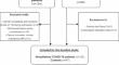
Methods: CORONIS (CORONavirus and Ischemic Stroke) was an observational cohort study in adult hospitalized patients for COVID-19 and controls without COVID-19, conducted between April 2021 and September 2022. Brain MRI was performed shortly after discharge and after 3 months. Outcomes included recent ischemic (DWI-positive) lesions, previous infarction, microbleeds, white matter hyperintensities (WMH) and intracerebral hemorrhage and were analysed with logistic regression to adjust for confounders.
Results: 125 patients with COVID-19 and 47 controls underwent brain MRI a median of 41.5 days after symptom onset. DWI-positive lesions were found in one patient (1%) and in one (2%) control, both clinically silent. WMH were more prevalent in patients (78%) than in controls (62%) (adjusted OR: 2.95 [95% CI: 1.07-8.57]), other cerebrovascular MRI markers did not differ. Prevalence of markers in ICU vs. non-ICU patients was similar. After three months, five patients (5%) had new cerebrovascular lesions, including DWI-positive lesions (1 patient, 1.0%), cerebral infarction (2 patients, 2.0%) and microbleeds (3 patients, 3.1%).
Conclusion: Overall, we found no higher prevalence of cerebrovascular markers in unselected hospitalized COVID-19 patients compared to controls. The few incident DWI-lesions were most likely to be explained by risk-factors of small vessel disease. In the general hospitalized COVID-19 population, COVID-19 shows limited impact on cerebrovascular MRI markers shortly after hospitalization.
期刊介绍:
Neuroradiology aims to provide state-of-the-art medical and scientific information in the fields of Neuroradiology, Neurosciences, Neurology, Psychiatry, Neurosurgery, and related medical specialities. Neuroradiology as the official Journal of the European Society of Neuroradiology receives submissions from all parts of the world and publishes peer-reviewed original research, comprehensive reviews, educational papers, opinion papers, and short reports on exceptional clinical observations and new technical developments in the field of Neuroimaging and Neurointervention. The journal has subsections for Diagnostic and Interventional Neuroradiology, Advanced Neuroimaging, Paediatric Neuroradiology, Head-Neck-ENT Radiology, Spine Neuroradiology, and for submissions from Japan. Neuroradiology aims to provide new knowledge about and insights into the function and pathology of the human nervous system that may help to better diagnose and treat nervous system diseases. Neuroradiology is a member of the Committee on Publication Ethics (COPE) and follows the COPE core practices. Neuroradiology prefers articles that are free of bias, self-critical regarding limitations, transparent and clear in describing study participants, methods, and statistics, and short in presenting results. Before peer-review all submissions are automatically checked by iThenticate to assess for potential overlap in prior publication.


 分享
分享
 求助内容:
求助内容: 应助结果提醒方式:
 扫码关注我们
