{"title":"基于锥形束计算机断层扫描的恒前磨牙牙根扩张评估:回顾性研究","authors":"Bahar Asheghi, Safoora Sahebi, Zeinab Rafiee, Maryam Zangooei Booshehri, Afsane Habibi","doi":"10.30476/dentjods.2023.98244.2067","DOIUrl":null,"url":null,"abstract":"<p><strong>Statement of the problem: </strong>As a developmental disorder characterized by an abnormal bend and angle in the longitudinal axis of the tooth root, dilaceration can cause complications in routine dental procedures such as endodontics, orthodontics, and surgical treatments.</p><p><strong>Purpose: </strong>The purpose of this study was to investigate the prevalence of dilaceration in maxillary and mandibular premolar teeth in a population of Shiraz city based on cone-beam computed tomography (CBCT).</p><p><strong>Materials and method: </strong>This is a retrospective cross-sectional study on 927 premolar teeth and 132 CBCT radiographs of patients obtained from four private radiology clinics in Shiraz (Iran). In this study, the presence, location, direction, and severity of dilaceration in premolar roots as well as its relationship with gender were investigated. Chi-square and Fisher tests were used to analyze the data.</p><p><strong>Results: </strong>The results showed that 17% of the studied 927 teeth had dilaceration. The prevalence of dilaceration was significantly higher in women than in men (20.3% vs. 13.6%, <i>p</i>= 0.005). The dilaceration rates were significantly higher in the mandibular first and second premolar teeth (31.6% and 26%, <i>p</i>= 0.002) than in the other teeth. In addition, the highest prevalence was in the distal direction with mild severity in the apical third of the root (<i>p</i>< 0.001).</p><p><strong>Conclusion: </strong>According to the results of this study, the prevalence of dilaceration was relatively high in mandibular premolar teeth especially in women.</p>","PeriodicalId":73702,"journal":{"name":"Journal of dentistry (Shiraz, Iran)","volume":"25 2","pages":"155-161"},"PeriodicalIF":0.0000,"publicationDate":"2024-06-01","publicationTypes":"Journal Article","fieldsOfStudy":null,"isOpenAccess":false,"openAccessPdf":"https://www.ncbi.nlm.nih.gov/pmc/articles/PMC11217063/pdf/","citationCount":"0","resultStr":"{\"title\":\"A Cone-Beam Computed Tomography-Based Evaluation of Root Dilaceration in Permanent Premolars: A Retrospective Study.\",\"authors\":\"Bahar Asheghi, Safoora Sahebi, Zeinab Rafiee, Maryam Zangooei Booshehri, Afsane Habibi\",\"doi\":\"10.30476/dentjods.2023.98244.2067\",\"DOIUrl\":null,\"url\":null,\"abstract\":\"<p><strong>Statement of the problem: </strong>As a developmental disorder characterized by an abnormal bend and angle in the longitudinal axis of the tooth root, dilaceration can cause complications in routine dental procedures such as endodontics, orthodontics, and surgical treatments.</p><p><strong>Purpose: </strong>The purpose of this study was to investigate the prevalence of dilaceration in maxillary and mandibular premolar teeth in a population of Shiraz city based on cone-beam computed tomography (CBCT).</p><p><strong>Materials and method: </strong>This is a retrospective cross-sectional study on 927 premolar teeth and 132 CBCT radiographs of patients obtained from four private radiology clinics in Shiraz (Iran). In this study, the presence, location, direction, and severity of dilaceration in premolar roots as well as its relationship with gender were investigated. Chi-square and Fisher tests were used to analyze the data.</p><p><strong>Results: </strong>The results showed that 17% of the studied 927 teeth had dilaceration. The prevalence of dilaceration was significantly higher in women than in men (20.3% vs. 13.6%, <i>p</i>= 0.005). The dilaceration rates were significantly higher in the mandibular first and second premolar teeth (31.6% and 26%, <i>p</i>= 0.002) than in the other teeth. In addition, the highest prevalence was in the distal direction with mild severity in the apical third of the root (<i>p</i>< 0.001).</p><p><strong>Conclusion: </strong>According to the results of this study, the prevalence of dilaceration was relatively high in mandibular premolar teeth especially in women.</p>\",\"PeriodicalId\":73702,\"journal\":{\"name\":\"Journal of dentistry (Shiraz, Iran)\",\"volume\":\"25 2\",\"pages\":\"155-161\"},\"PeriodicalIF\":0.0000,\"publicationDate\":\"2024-06-01\",\"publicationTypes\":\"Journal Article\",\"fieldsOfStudy\":null,\"isOpenAccess\":false,\"openAccessPdf\":\"https://www.ncbi.nlm.nih.gov/pmc/articles/PMC11217063/pdf/\",\"citationCount\":\"0\",\"resultStr\":null,\"platform\":\"Semanticscholar\",\"paperid\":null,\"PeriodicalName\":\"Journal of dentistry (Shiraz, Iran)\",\"FirstCategoryId\":\"1085\",\"ListUrlMain\":\"https://doi.org/10.30476/dentjods.2023.98244.2067\",\"RegionNum\":0,\"RegionCategory\":null,\"ArticlePicture\":[],\"TitleCN\":null,\"AbstractTextCN\":null,\"PMCID\":null,\"EPubDate\":\"\",\"PubModel\":\"\",\"JCR\":\"\",\"JCRName\":\"\",\"Score\":null,\"Total\":0}","platform":"Semanticscholar","paperid":null,"PeriodicalName":"Journal of dentistry (Shiraz, Iran)","FirstCategoryId":"1085","ListUrlMain":"https://doi.org/10.30476/dentjods.2023.98244.2067","RegionNum":0,"RegionCategory":null,"ArticlePicture":[],"TitleCN":null,"AbstractTextCN":null,"PMCID":null,"EPubDate":"","PubModel":"","JCR":"","JCRName":"","Score":null,"Total":0}
A Cone-Beam Computed Tomography-Based Evaluation of Root Dilaceration in Permanent Premolars: A Retrospective Study.
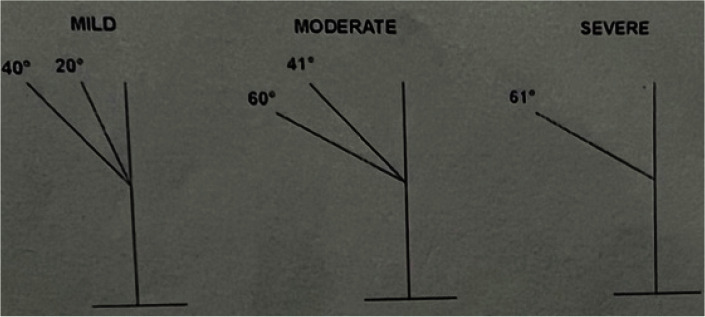
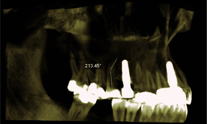
Statement of the problem: As a developmental disorder characterized by an abnormal bend and angle in the longitudinal axis of the tooth root, dilaceration can cause complications in routine dental procedures such as endodontics, orthodontics, and surgical treatments.
Purpose: The purpose of this study was to investigate the prevalence of dilaceration in maxillary and mandibular premolar teeth in a population of Shiraz city based on cone-beam computed tomography (CBCT).
Materials and method: This is a retrospective cross-sectional study on 927 premolar teeth and 132 CBCT radiographs of patients obtained from four private radiology clinics in Shiraz (Iran). In this study, the presence, location, direction, and severity of dilaceration in premolar roots as well as its relationship with gender were investigated. Chi-square and Fisher tests were used to analyze the data.
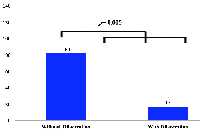
Results: The results showed that 17% of the studied 927 teeth had dilaceration. The prevalence of dilaceration was significantly higher in women than in men (20.3% vs. 13.6%, p= 0.005). The dilaceration rates were significantly higher in the mandibular first and second premolar teeth (31.6% and 26%, p= 0.002) than in the other teeth. In addition, the highest prevalence was in the distal direction with mild severity in the apical third of the root (p< 0.001).
Conclusion: According to the results of this study, the prevalence of dilaceration was relatively high in mandibular premolar teeth especially in women.


 分享
分享

 求助内容:
求助内容: 应助结果提醒方式:
 扫码关注我们
