Anu Singh, Jagruti Patil, Sitaram G. Ghogale, Nilesh Deshpande, Karishma Girase, Navami Shetye, Sweta Rajpal, Gaurav Chatterjee, Nikhil Patkar, Disha Jain, Sridhar Epari, Tanuja Shet, Sumeet Gujral, Papagudi G. Subramanian, Prashant R. Tembhare
{"title":"白细胞相关免疫球蛋白样受体-1(CD305)在流式细胞术检测 B 细胞非霍奇金淋巴瘤骨髓微小受累情况中的实用性","authors":"Anu Singh, Jagruti Patil, Sitaram G. Ghogale, Nilesh Deshpande, Karishma Girase, Navami Shetye, Sweta Rajpal, Gaurav Chatterjee, Nikhil Patkar, Disha Jain, Sridhar Epari, Tanuja Shet, Sumeet Gujral, Papagudi G. Subramanian, Prashant R. Tembhare","doi":"10.1002/cyto.b.22193","DOIUrl":null,"url":null,"abstract":"<p>Multicolor flow cytometry (MFC) is crucial in detecting occult or minimal bone marrow (BM) involvement by non-Hodgkin lymphomas (NHL), which may not be detected using trephine biopsy or imaging studies. Detection of low-level BM involvement can be challenging without definite immunophenotypic aberrancies. We studied the utility of CD305 in MFC detection of minimal BM involvement by B-NHL, especially in the absence of aberrancies by commonly used markers. The study included 1084 consecutive BM samples submitted for the staging of B-NHLs (excluding CLL) over two years. Samples were studied for morphological, immunophenotypic, and histopathological assessment. MFC studies were performed using 10–13 color MFC, including CD305-antibody (clone, DX26<b>)</b>. Minimal BM involvement was defined with a cutoff of ≤10% lymphoma cells in viable cells on MFC assessment. Of 1084, 148 samples revealed overt morphological involvement by B-NHL and were excluded from analysis. BM samples of 172/936 patients were morphologically negative but revealed involvement using MFC independently. Corresponding trephine biopsy involvement was detected in only 79/172 (45.9%) patients. On MFC, 23/172 samples showed BM involvement with >10% lymphoma cells, and 149/172 (86.6%) samples revealed minimal involvement. In 54/149 (36.24%) samples, lymphoma cells were detected only with aberrant loss of CD305 expression. In 78 of the remaining 95 samples (82.1%), it provided an immunophenotypic aberrancy addition to other markers and supported the results. CD305 is a highly useful marker in the flow cytometric assessment of minimal BM involvement by B-NHL. MFC is a superior modality to trephine biopsy in detecting low-level BM involvement.</p>","PeriodicalId":10883,"journal":{"name":"Cytometry Part B: Clinical Cytometry","volume":"106 5","pages":"359-369"},"PeriodicalIF":2.7000,"publicationDate":"2024-06-21","publicationTypes":"Journal Article","fieldsOfStudy":null,"isOpenAccess":false,"openAccessPdf":"https://onlinelibrary.wiley.com/doi/epdf/10.1002/cyto.b.22193","citationCount":"0","resultStr":"{\"title\":\"Utility of leukocyte-associated immunoglobulin-like receptor-1 (CD305) in flow cytometric detection of minimal bone marrow involvement by B-cell non-Hodgkin lymphoma\",\"authors\":\"Anu Singh, Jagruti Patil, Sitaram G. Ghogale, Nilesh Deshpande, Karishma Girase, Navami Shetye, Sweta Rajpal, Gaurav Chatterjee, Nikhil Patkar, Disha Jain, Sridhar Epari, Tanuja Shet, Sumeet Gujral, Papagudi G. Subramanian, Prashant R. Tembhare\",\"doi\":\"10.1002/cyto.b.22193\",\"DOIUrl\":null,\"url\":null,\"abstract\":\"<p>Multicolor flow cytometry (MFC) is crucial in detecting occult or minimal bone marrow (BM) involvement by non-Hodgkin lymphomas (NHL), which may not be detected using trephine biopsy or imaging studies. Detection of low-level BM involvement can be challenging without definite immunophenotypic aberrancies. We studied the utility of CD305 in MFC detection of minimal BM involvement by B-NHL, especially in the absence of aberrancies by commonly used markers. The study included 1084 consecutive BM samples submitted for the staging of B-NHLs (excluding CLL) over two years. Samples were studied for morphological, immunophenotypic, and histopathological assessment. MFC studies were performed using 10–13 color MFC, including CD305-antibody (clone, DX26<b>)</b>. Minimal BM involvement was defined with a cutoff of ≤10% lymphoma cells in viable cells on MFC assessment. Of 1084, 148 samples revealed overt morphological involvement by B-NHL and were excluded from analysis. BM samples of 172/936 patients were morphologically negative but revealed involvement using MFC independently. Corresponding trephine biopsy involvement was detected in only 79/172 (45.9%) patients. On MFC, 23/172 samples showed BM involvement with >10% lymphoma cells, and 149/172 (86.6%) samples revealed minimal involvement. In 54/149 (36.24%) samples, lymphoma cells were detected only with aberrant loss of CD305 expression. In 78 of the remaining 95 samples (82.1%), it provided an immunophenotypic aberrancy addition to other markers and supported the results. CD305 is a highly useful marker in the flow cytometric assessment of minimal BM involvement by B-NHL. MFC is a superior modality to trephine biopsy in detecting low-level BM involvement.</p>\",\"PeriodicalId\":10883,\"journal\":{\"name\":\"Cytometry Part B: Clinical Cytometry\",\"volume\":\"106 5\",\"pages\":\"359-369\"},\"PeriodicalIF\":2.7000,\"publicationDate\":\"2024-06-21\",\"publicationTypes\":\"Journal Article\",\"fieldsOfStudy\":null,\"isOpenAccess\":false,\"openAccessPdf\":\"https://onlinelibrary.wiley.com/doi/epdf/10.1002/cyto.b.22193\",\"citationCount\":\"0\",\"resultStr\":null,\"platform\":\"Semanticscholar\",\"paperid\":null,\"PeriodicalName\":\"Cytometry Part B: Clinical Cytometry\",\"FirstCategoryId\":\"3\",\"ListUrlMain\":\"https://onlinelibrary.wiley.com/doi/10.1002/cyto.b.22193\",\"RegionNum\":3,\"RegionCategory\":\"医学\",\"ArticlePicture\":[],\"TitleCN\":null,\"AbstractTextCN\":null,\"PMCID\":null,\"EPubDate\":\"\",\"PubModel\":\"\",\"JCR\":\"Q3\",\"JCRName\":\"MEDICAL LABORATORY TECHNOLOGY\",\"Score\":null,\"Total\":0}","platform":"Semanticscholar","paperid":null,"PeriodicalName":"Cytometry Part B: Clinical Cytometry","FirstCategoryId":"3","ListUrlMain":"https://onlinelibrary.wiley.com/doi/10.1002/cyto.b.22193","RegionNum":3,"RegionCategory":"医学","ArticlePicture":[],"TitleCN":null,"AbstractTextCN":null,"PMCID":null,"EPubDate":"","PubModel":"","JCR":"Q3","JCRName":"MEDICAL LABORATORY TECHNOLOGY","Score":null,"Total":0}
Utility of leukocyte-associated immunoglobulin-like receptor-1 (CD305) in flow cytometric detection of minimal bone marrow involvement by B-cell non-Hodgkin lymphoma
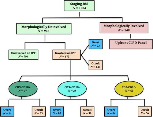
Multicolor flow cytometry (MFC) is crucial in detecting occult or minimal bone marrow (BM) involvement by non-Hodgkin lymphomas (NHL), which may not be detected using trephine biopsy or imaging studies. Detection of low-level BM involvement can be challenging without definite immunophenotypic aberrancies. We studied the utility of CD305 in MFC detection of minimal BM involvement by B-NHL, especially in the absence of aberrancies by commonly used markers. The study included 1084 consecutive BM samples submitted for the staging of B-NHLs (excluding CLL) over two years. Samples were studied for morphological, immunophenotypic, and histopathological assessment. MFC studies were performed using 10–13 color MFC, including CD305-antibody (clone, DX26). Minimal BM involvement was defined with a cutoff of ≤10% lymphoma cells in viable cells on MFC assessment. Of 1084, 148 samples revealed overt morphological involvement by B-NHL and were excluded from analysis. BM samples of 172/936 patients were morphologically negative but revealed involvement using MFC independently. Corresponding trephine biopsy involvement was detected in only 79/172 (45.9%) patients. On MFC, 23/172 samples showed BM involvement with >10% lymphoma cells, and 149/172 (86.6%) samples revealed minimal involvement. In 54/149 (36.24%) samples, lymphoma cells were detected only with aberrant loss of CD305 expression. In 78 of the remaining 95 samples (82.1%), it provided an immunophenotypic aberrancy addition to other markers and supported the results. CD305 is a highly useful marker in the flow cytometric assessment of minimal BM involvement by B-NHL. MFC is a superior modality to trephine biopsy in detecting low-level BM involvement.
期刊介绍:
Cytometry Part B: Clinical Cytometry features original research reports, in-depth reviews and special issues that directly relate to and palpably impact clinical flow, mass and image-based cytometry. These may include clinical and translational investigations important in the diagnostic, prognostic and therapeutic management of patients. Thus, we welcome research papers from various disciplines related [but not limited to] hematopathologists, hematologists, immunologists and cell biologists with clinically relevant and innovative studies investigating individual-cell analytics and/or separations. In addition to the types of papers indicated above, we also welcome Letters to the Editor, describing case reports or important medical or technical topics relevant to our readership without the length and depth of a full original report.


 分享
分享
 求助内容:
求助内容: 应助结果提醒方式:
 扫码关注我们
