R. R. Sharipov, I. A. Tarzhanov, A. A. Zgodova, Z. V. Bakaeva, A. M. Surin
{"title":"使用基于阿塞丹的荧光探针监测原代神经元培养物中硫化氢的可行性","authors":"R. R. Sharipov, I. A. Tarzhanov, A. A. Zgodova, Z. V. Bakaeva, A. M. Surin","doi":"10.1134/S1990747824700119","DOIUrl":null,"url":null,"abstract":"<p>Hydrogen sulfide (H<sub>2</sub>S), which under physiological conditions exists in cells mainly in the form of the HS<sup>–</sup> anion, is considered as a gaseous transmitter of inter- and intracellular signals along with nitrogen monoxide and carbon monoxide. Analysis of the dynamics of H<sub>2</sub>S content in living cells is impossible without the creation of sensitive and specific probes. The group of K.H. Ahn synthesized several acedan-based compounds, which in the presence of H<sub>2</sub>S attached a sulfhydryl group, forming fluorescent carbocyclic compounds. According to the spectral characteristics and reaction rate with H<sub>2</sub>S, the optimal substance was P3, which forms the carbocyclic compound csP3 with the same large Stokes shift as P3 (approx. 130 nm) and has a brighter fluorescence. In this work, we tested the suitability of csP3 for recording changes in H<sub>2</sub>S in solutions simulating the minimum salt composition of the intracellular medium, as well as in cells of primary neuronal culture from the rat cerebral cortex. It was found that the fluorescence intensity of csP3, which was formed when Na<sub>2</sub>S (H<sub>2</sub>S donor, 100 and 300 µM) was added to the P3 solution, differed for solutions corresponding in salt composition to the extracellular medium and cytosol. In both cases, fluorescence increased in the presence of bicarbonate (NaHCO<sub>3</sub>, 10 mM). A decrease in the polarity of solutions due to the addition of dimethyl sulfoxide (30% by volume) shifted the emission by approx. 10 nm to the shorter wavelength region and doubled the intensity. Glutamate (10 µM, in the presence of 10 µM of glycine, 0 Mg<sup>2+</sup>) increased the fluorescence of the probe, but only in those neurons in which delayed deregulation of calcium homeostasis did not occur. The addition of P3 or csP3 to the cell culture caused a rapid increase in the fluorescent signal, which was replaced by a slow signal growth after 3–5 min. It was concluded that the product of the reaction of P3 with H<sub>2</sub>S was sensitive to changes in the salt composition of the intracellular medium and could be redistributed in cells between an aqueous and more hydrophobic environment. These circumstances made it difficult to interpret the growth of P3 fluorescence in cells as a quantitative indicator of the presence of H<sub>2</sub>S and required additional studies of the properties of this and structurally related H<sub>2</sub>S probes.</p>","PeriodicalId":484,"journal":{"name":"Biochemistry (Moscow), Supplement Series A: Membrane and Cell Biology","volume":"18 2","pages":"90 - 99"},"PeriodicalIF":0.6000,"publicationDate":"2024-06-25","publicationTypes":"Journal Article","fieldsOfStudy":null,"isOpenAccess":false,"openAccessPdf":"","citationCount":"0","resultStr":"{\"title\":\"On the Feasibility of Using an Acedan-Based Fluorescent Probe to Monitor Hydrogen Sulfide in Primary Neuronal Cultures\",\"authors\":\"R. R. Sharipov, I. A. Tarzhanov, A. A. Zgodova, Z. V. Bakaeva, A. M. Surin\",\"doi\":\"10.1134/S1990747824700119\",\"DOIUrl\":null,\"url\":null,\"abstract\":\"<p>Hydrogen sulfide (H<sub>2</sub>S), which under physiological conditions exists in cells mainly in the form of the HS<sup>–</sup> anion, is considered as a gaseous transmitter of inter- and intracellular signals along with nitrogen monoxide and carbon monoxide. Analysis of the dynamics of H<sub>2</sub>S content in living cells is impossible without the creation of sensitive and specific probes. The group of K.H. Ahn synthesized several acedan-based compounds, which in the presence of H<sub>2</sub>S attached a sulfhydryl group, forming fluorescent carbocyclic compounds. According to the spectral characteristics and reaction rate with H<sub>2</sub>S, the optimal substance was P3, which forms the carbocyclic compound csP3 with the same large Stokes shift as P3 (approx. 130 nm) and has a brighter fluorescence. In this work, we tested the suitability of csP3 for recording changes in H<sub>2</sub>S in solutions simulating the minimum salt composition of the intracellular medium, as well as in cells of primary neuronal culture from the rat cerebral cortex. It was found that the fluorescence intensity of csP3, which was formed when Na<sub>2</sub>S (H<sub>2</sub>S donor, 100 and 300 µM) was added to the P3 solution, differed for solutions corresponding in salt composition to the extracellular medium and cytosol. In both cases, fluorescence increased in the presence of bicarbonate (NaHCO<sub>3</sub>, 10 mM). A decrease in the polarity of solutions due to the addition of dimethyl sulfoxide (30% by volume) shifted the emission by approx. 10 nm to the shorter wavelength region and doubled the intensity. Glutamate (10 µM, in the presence of 10 µM of glycine, 0 Mg<sup>2+</sup>) increased the fluorescence of the probe, but only in those neurons in which delayed deregulation of calcium homeostasis did not occur. The addition of P3 or csP3 to the cell culture caused a rapid increase in the fluorescent signal, which was replaced by a slow signal growth after 3–5 min. It was concluded that the product of the reaction of P3 with H<sub>2</sub>S was sensitive to changes in the salt composition of the intracellular medium and could be redistributed in cells between an aqueous and more hydrophobic environment. These circumstances made it difficult to interpret the growth of P3 fluorescence in cells as a quantitative indicator of the presence of H<sub>2</sub>S and required additional studies of the properties of this and structurally related H<sub>2</sub>S probes.</p>\",\"PeriodicalId\":484,\"journal\":{\"name\":\"Biochemistry (Moscow), Supplement Series A: Membrane and Cell Biology\",\"volume\":\"18 2\",\"pages\":\"90 - 99\"},\"PeriodicalIF\":0.6000,\"publicationDate\":\"2024-06-25\",\"publicationTypes\":\"Journal Article\",\"fieldsOfStudy\":null,\"isOpenAccess\":false,\"openAccessPdf\":\"\",\"citationCount\":\"0\",\"resultStr\":null,\"platform\":\"Semanticscholar\",\"paperid\":null,\"PeriodicalName\":\"Biochemistry (Moscow), Supplement Series A: Membrane and Cell Biology\",\"FirstCategoryId\":\"2\",\"ListUrlMain\":\"https://link.springer.com/article/10.1134/S1990747824700119\",\"RegionNum\":0,\"RegionCategory\":null,\"ArticlePicture\":[],\"TitleCN\":null,\"AbstractTextCN\":null,\"PMCID\":null,\"EPubDate\":\"\",\"PubModel\":\"\",\"JCR\":\"Q4\",\"JCRName\":\"CELL BIOLOGY\",\"Score\":null,\"Total\":0}","platform":"Semanticscholar","paperid":null,"PeriodicalName":"Biochemistry (Moscow), Supplement Series A: Membrane and Cell Biology","FirstCategoryId":"2","ListUrlMain":"https://link.springer.com/article/10.1134/S1990747824700119","RegionNum":0,"RegionCategory":null,"ArticlePicture":[],"TitleCN":null,"AbstractTextCN":null,"PMCID":null,"EPubDate":"","PubModel":"","JCR":"Q4","JCRName":"CELL BIOLOGY","Score":null,"Total":0}
On the Feasibility of Using an Acedan-Based Fluorescent Probe to Monitor Hydrogen Sulfide in Primary Neuronal Cultures
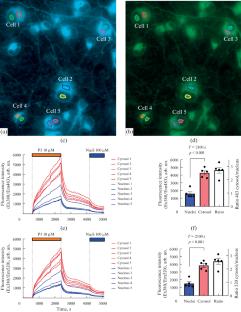
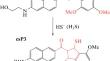
Hydrogen sulfide (H2S), which under physiological conditions exists in cells mainly in the form of the HS– anion, is considered as a gaseous transmitter of inter- and intracellular signals along with nitrogen monoxide and carbon monoxide. Analysis of the dynamics of H2S content in living cells is impossible without the creation of sensitive and specific probes. The group of K.H. Ahn synthesized several acedan-based compounds, which in the presence of H2S attached a sulfhydryl group, forming fluorescent carbocyclic compounds. According to the spectral characteristics and reaction rate with H2S, the optimal substance was P3, which forms the carbocyclic compound csP3 with the same large Stokes shift as P3 (approx. 130 nm) and has a brighter fluorescence. In this work, we tested the suitability of csP3 for recording changes in H2S in solutions simulating the minimum salt composition of the intracellular medium, as well as in cells of primary neuronal culture from the rat cerebral cortex. It was found that the fluorescence intensity of csP3, which was formed when Na2S (H2S donor, 100 and 300 µM) was added to the P3 solution, differed for solutions corresponding in salt composition to the extracellular medium and cytosol. In both cases, fluorescence increased in the presence of bicarbonate (NaHCO3, 10 mM). A decrease in the polarity of solutions due to the addition of dimethyl sulfoxide (30% by volume) shifted the emission by approx. 10 nm to the shorter wavelength region and doubled the intensity. Glutamate (10 µM, in the presence of 10 µM of glycine, 0 Mg2+) increased the fluorescence of the probe, but only in those neurons in which delayed deregulation of calcium homeostasis did not occur. The addition of P3 or csP3 to the cell culture caused a rapid increase in the fluorescent signal, which was replaced by a slow signal growth after 3–5 min. It was concluded that the product of the reaction of P3 with H2S was sensitive to changes in the salt composition of the intracellular medium and could be redistributed in cells between an aqueous and more hydrophobic environment. These circumstances made it difficult to interpret the growth of P3 fluorescence in cells as a quantitative indicator of the presence of H2S and required additional studies of the properties of this and structurally related H2S probes.
期刊介绍:
Biochemistry (Moscow), Supplement Series A: Membrane and Cell Biology is an international peer reviewed journal that publishes original articles on physical, chemical, and molecular mechanisms that underlie basic properties of biological membranes and mediate membrane-related cellular functions. The primary topics of the journal are membrane structure, mechanisms of membrane transport, bioenergetics and photobiology, intracellular signaling as well as membrane aspects of cell biology, immunology, and medicine. The journal is multidisciplinary and gives preference to those articles that employ a variety of experimental approaches, basically in biophysics but also in biochemistry, cytology, and molecular biology. The journal publishes articles that strive for unveiling membrane and cellular functions through innovative theoretical models and computer simulations.


 分享
分享
 求助内容:
求助内容: 应助结果提醒方式:
 扫码关注我们
