{"title":"研究纳米和微量氧化镍不同给药途径对大鼠肾脏结构、细胞凋亡标志物、氧化应激和组织病理学的影响。","authors":"Hatice Karaboduk, Caglar Adiguzel, Fatma Gokce Apaydin, Suna Kalender, Yusuf Kalender","doi":"10.1007/s10735-024-10221-5","DOIUrl":null,"url":null,"abstract":"<div><p>Although the production and use of nickel oxide nanoparticles (NiONP) are widespread, environmental and public health problems are associated with it. The kidney is the primary organ in excretion and is among the target organs in nanoparticle toxicity. This study aimed to compare the renal toxicity of nickel oxide (NiO) microparticles and nickel oxide nanoparticles by different routes of administration, such as oral, intraperitoneal (IP), and intravenous (IV). Seven groups were formed, with 42 male rats and six animals in each group. NiO oral (150 mg/kg), NiO IP (20 mg/kg), NiO IV (1 mg/kg), NiONP oral (150 mg/kg), NiONP IP (20 mg/kg), and NiONP IV (1 mg/kg) was administered for 21 days. After NiO and NiONP administration, a decrease in antioxidant activities and an increase in lipid peroxidation occurred in the kidney tissue of rats. Increased kidney urea, uric acid, and creatinine levels were observed. Inhibition of acetylcholinesterase activity and an increase in interleukin 1 beta were detected. Apoptotic markers, Bax, caspase-3, and p53 up-regulation and Bcl-2 down-regulation were observed. In addition, histopathological changes occurred in the kidney tissue. In general, it was observed that nickel oxide microparticles and nickel oxide nanoparticles cause inflammation by causing oxidative stress in the kidney tissue, and NiONP IV administration is more effective in renal toxicity.</p><h3>Graphical abstract</h3>\n<div><figure><div><div><picture><source><img></source></picture></div></div></figure></div></div>","PeriodicalId":650,"journal":{"name":"Journal of Molecular Histology","volume":"55 5","pages":"675 - 686"},"PeriodicalIF":2.2000,"publicationDate":"2024-07-11","publicationTypes":"Journal Article","fieldsOfStudy":null,"isOpenAccess":false,"openAccessPdf":"","citationCount":"0","resultStr":"{\"title\":\"Investigating the impact of different routes of nano and micro nickel oxide administration on rat kidney architecture, apoptosis markers, oxidative stress, and histopathology\",\"authors\":\"Hatice Karaboduk, Caglar Adiguzel, Fatma Gokce Apaydin, Suna Kalender, Yusuf Kalender\",\"doi\":\"10.1007/s10735-024-10221-5\",\"DOIUrl\":null,\"url\":null,\"abstract\":\"<div><p>Although the production and use of nickel oxide nanoparticles (NiONP) are widespread, environmental and public health problems are associated with it. The kidney is the primary organ in excretion and is among the target organs in nanoparticle toxicity. This study aimed to compare the renal toxicity of nickel oxide (NiO) microparticles and nickel oxide nanoparticles by different routes of administration, such as oral, intraperitoneal (IP), and intravenous (IV). Seven groups were formed, with 42 male rats and six animals in each group. NiO oral (150 mg/kg), NiO IP (20 mg/kg), NiO IV (1 mg/kg), NiONP oral (150 mg/kg), NiONP IP (20 mg/kg), and NiONP IV (1 mg/kg) was administered for 21 days. After NiO and NiONP administration, a decrease in antioxidant activities and an increase in lipid peroxidation occurred in the kidney tissue of rats. Increased kidney urea, uric acid, and creatinine levels were observed. Inhibition of acetylcholinesterase activity and an increase in interleukin 1 beta were detected. Apoptotic markers, Bax, caspase-3, and p53 up-regulation and Bcl-2 down-regulation were observed. In addition, histopathological changes occurred in the kidney tissue. In general, it was observed that nickel oxide microparticles and nickel oxide nanoparticles cause inflammation by causing oxidative stress in the kidney tissue, and NiONP IV administration is more effective in renal toxicity.</p><h3>Graphical abstract</h3>\\n<div><figure><div><div><picture><source><img></source></picture></div></div></figure></div></div>\",\"PeriodicalId\":650,\"journal\":{\"name\":\"Journal of Molecular Histology\",\"volume\":\"55 5\",\"pages\":\"675 - 686\"},\"PeriodicalIF\":2.2000,\"publicationDate\":\"2024-07-11\",\"publicationTypes\":\"Journal Article\",\"fieldsOfStudy\":null,\"isOpenAccess\":false,\"openAccessPdf\":\"\",\"citationCount\":\"0\",\"resultStr\":null,\"platform\":\"Semanticscholar\",\"paperid\":null,\"PeriodicalName\":\"Journal of Molecular Histology\",\"FirstCategoryId\":\"99\",\"ListUrlMain\":\"https://link.springer.com/article/10.1007/s10735-024-10221-5\",\"RegionNum\":4,\"RegionCategory\":\"生物学\",\"ArticlePicture\":[],\"TitleCN\":null,\"AbstractTextCN\":null,\"PMCID\":null,\"EPubDate\":\"\",\"PubModel\":\"\",\"JCR\":\"Q3\",\"JCRName\":\"CELL BIOLOGY\",\"Score\":null,\"Total\":0}","platform":"Semanticscholar","paperid":null,"PeriodicalName":"Journal of Molecular Histology","FirstCategoryId":"99","ListUrlMain":"https://link.springer.com/article/10.1007/s10735-024-10221-5","RegionNum":4,"RegionCategory":"生物学","ArticlePicture":[],"TitleCN":null,"AbstractTextCN":null,"PMCID":null,"EPubDate":"","PubModel":"","JCR":"Q3","JCRName":"CELL BIOLOGY","Score":null,"Total":0}
Investigating the impact of different routes of nano and micro nickel oxide administration on rat kidney architecture, apoptosis markers, oxidative stress, and histopathology
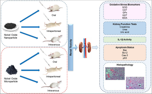
Although the production and use of nickel oxide nanoparticles (NiONP) are widespread, environmental and public health problems are associated with it. The kidney is the primary organ in excretion and is among the target organs in nanoparticle toxicity. This study aimed to compare the renal toxicity of nickel oxide (NiO) microparticles and nickel oxide nanoparticles by different routes of administration, such as oral, intraperitoneal (IP), and intravenous (IV). Seven groups were formed, with 42 male rats and six animals in each group. NiO oral (150 mg/kg), NiO IP (20 mg/kg), NiO IV (1 mg/kg), NiONP oral (150 mg/kg), NiONP IP (20 mg/kg), and NiONP IV (1 mg/kg) was administered for 21 days. After NiO and NiONP administration, a decrease in antioxidant activities and an increase in lipid peroxidation occurred in the kidney tissue of rats. Increased kidney urea, uric acid, and creatinine levels were observed. Inhibition of acetylcholinesterase activity and an increase in interleukin 1 beta were detected. Apoptotic markers, Bax, caspase-3, and p53 up-regulation and Bcl-2 down-regulation were observed. In addition, histopathological changes occurred in the kidney tissue. In general, it was observed that nickel oxide microparticles and nickel oxide nanoparticles cause inflammation by causing oxidative stress in the kidney tissue, and NiONP IV administration is more effective in renal toxicity.
期刊介绍:
The Journal of Molecular Histology publishes results of original research on the localization and expression of molecules in animal cells, tissues and organs. Coverage includes studies describing novel cellular or ultrastructural distributions of molecules which provide insight into biochemical or physiological function, development, histologic structure and disease processes.
Major research themes of particular interest include:
- Cell-Cell and Cell-Matrix Interactions;
- Connective Tissues;
- Development and Disease;
- Neuroscience.
Please note that the Journal of Molecular Histology does not consider manuscripts dealing with the application of immunological or other probes on non-standard laboratory animal models unless the results are clearly of significant and general biological importance.
The Journal of Molecular Histology publishes full-length original research papers, review articles, short communications and letters to the editors. All manuscripts are typically reviewed by two independent referees. The Journal of Molecular Histology is a continuation of The Histochemical Journal.


 分享
分享
 求助内容:
求助内容: 应助结果提醒方式:
 扫码关注我们
