Lingui Gu, Hualin Chen, Mingjiang Sun, Yihao Chen, Qinglei Shi, Jianbo Chang, Junji Wei, Wenbin Ma, Xinjie Bao, Renzhi Wang
{"title":"揭示脑出血的动态免疫景观:单细胞和空间转录组特征分析的启示。","authors":"Lingui Gu, Hualin Chen, Mingjiang Sun, Yihao Chen, Qinglei Shi, Jianbo Chang, Junji Wei, Wenbin Ma, Xinjie Bao, Renzhi Wang","doi":"10.1002/mco2.635","DOIUrl":null,"url":null,"abstract":"<p>Intracerebral hemorrhage (ICH) poses a formidable challenge in stroke management, with limited therapeutic options, particularly in the realm of immune-targeted interventions. Clinical trials targeting immune responses post-ICH have encountered setbacks, potentially attributable to the substantial cellular heterogeneity and intricate intercellular networks within the brain. Here, we present a pioneering investigation utilizing single-cell RNA sequencing and spatial transcriptome profiling at hyperacute (1 h), acute (24 h), and subacute (7 days) intervals post-ICH, aimed at unraveling the dynamic immunological landscape and spatial distributions within the cerebral tissue. Our comprehensive analysis revealed distinct cell differentiation patterns among myeloid and lymphocyte populations, along with delineated spatial distributions across various brain regions. Notably, we identified a subset of lymphocytes characterized by the expression of Spp1 and Lyz2, termed macrophage-associated lymphocytes, which exhibited close interactions with myeloid cells. Specifically, we observed prominent interactions between Lgmn+Macro-T cells and microglia through the spp1–cd44 pathway during the acute phase post-ICH in the choroid plexus. These findings represent a significant advancement in our understanding of immune cell dynamics at single-cell resolution across distinct post-ICH time points, thereby laying the groundwork for exploring critical temporal windows and informing the development of targeted therapeutic strategies.</p>","PeriodicalId":94133,"journal":{"name":"MedComm","volume":"5 7","pages":""},"PeriodicalIF":10.7000,"publicationDate":"2024-07-10","publicationTypes":"Journal Article","fieldsOfStudy":null,"isOpenAccess":false,"openAccessPdf":"https://www.ncbi.nlm.nih.gov/pmc/articles/PMC11233862/pdf/","citationCount":"0","resultStr":"{\"title\":\"Unraveling dynamic immunological landscapes in intracerebral hemorrhage: insights from single-cell and spatial transcriptomic profiling\",\"authors\":\"Lingui Gu, Hualin Chen, Mingjiang Sun, Yihao Chen, Qinglei Shi, Jianbo Chang, Junji Wei, Wenbin Ma, Xinjie Bao, Renzhi Wang\",\"doi\":\"10.1002/mco2.635\",\"DOIUrl\":null,\"url\":null,\"abstract\":\"<p>Intracerebral hemorrhage (ICH) poses a formidable challenge in stroke management, with limited therapeutic options, particularly in the realm of immune-targeted interventions. Clinical trials targeting immune responses post-ICH have encountered setbacks, potentially attributable to the substantial cellular heterogeneity and intricate intercellular networks within the brain. Here, we present a pioneering investigation utilizing single-cell RNA sequencing and spatial transcriptome profiling at hyperacute (1 h), acute (24 h), and subacute (7 days) intervals post-ICH, aimed at unraveling the dynamic immunological landscape and spatial distributions within the cerebral tissue. Our comprehensive analysis revealed distinct cell differentiation patterns among myeloid and lymphocyte populations, along with delineated spatial distributions across various brain regions. Notably, we identified a subset of lymphocytes characterized by the expression of Spp1 and Lyz2, termed macrophage-associated lymphocytes, which exhibited close interactions with myeloid cells. Specifically, we observed prominent interactions between Lgmn+Macro-T cells and microglia through the spp1–cd44 pathway during the acute phase post-ICH in the choroid plexus. These findings represent a significant advancement in our understanding of immune cell dynamics at single-cell resolution across distinct post-ICH time points, thereby laying the groundwork for exploring critical temporal windows and informing the development of targeted therapeutic strategies.</p>\",\"PeriodicalId\":94133,\"journal\":{\"name\":\"MedComm\",\"volume\":\"5 7\",\"pages\":\"\"},\"PeriodicalIF\":10.7000,\"publicationDate\":\"2024-07-10\",\"publicationTypes\":\"Journal Article\",\"fieldsOfStudy\":null,\"isOpenAccess\":false,\"openAccessPdf\":\"https://www.ncbi.nlm.nih.gov/pmc/articles/PMC11233862/pdf/\",\"citationCount\":\"0\",\"resultStr\":null,\"platform\":\"Semanticscholar\",\"paperid\":null,\"PeriodicalName\":\"MedComm\",\"FirstCategoryId\":\"1085\",\"ListUrlMain\":\"https://onlinelibrary.wiley.com/doi/10.1002/mco2.635\",\"RegionNum\":0,\"RegionCategory\":null,\"ArticlePicture\":[],\"TitleCN\":null,\"AbstractTextCN\":null,\"PMCID\":null,\"EPubDate\":\"\",\"PubModel\":\"\",\"JCR\":\"Q1\",\"JCRName\":\"MEDICINE, RESEARCH & EXPERIMENTAL\",\"Score\":null,\"Total\":0}","platform":"Semanticscholar","paperid":null,"PeriodicalName":"MedComm","FirstCategoryId":"1085","ListUrlMain":"https://onlinelibrary.wiley.com/doi/10.1002/mco2.635","RegionNum":0,"RegionCategory":null,"ArticlePicture":[],"TitleCN":null,"AbstractTextCN":null,"PMCID":null,"EPubDate":"","PubModel":"","JCR":"Q1","JCRName":"MEDICINE, RESEARCH & EXPERIMENTAL","Score":null,"Total":0}
引用次数: 0
摘要
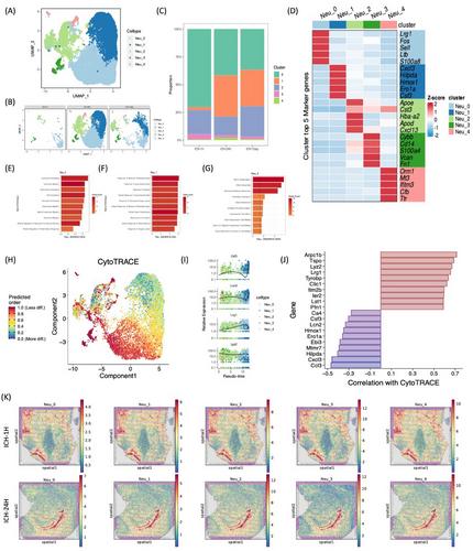
脑内出血(ICH)给中风治疗带来了严峻的挑战,因为治疗方案有限,尤其是在免疫靶向干预领域。针对 ICH 后免疫反应的临床试验遇到了挫折,这可能归因于脑内大量的细胞异质性和错综复杂的细胞间网络。在这里,我们利用单细胞 RNA 测序和空间转录组图谱对ICH 后的超急性期(1 小时)、急性期(24 小时)和亚急性期(7 天)进行了开创性的研究,旨在揭示脑组织内的动态免疫格局和空间分布。我们的综合分析揭示了髓细胞和淋巴细胞群之间不同的细胞分化模式,以及在不同脑区的空间分布。值得注意的是,我们发现了以表达 Spp1 和 Lyz2 为特征的淋巴细胞亚群,即巨噬细胞相关淋巴细胞,它们与髓系细胞表现出密切的相互作用。具体来说,我们观察到 Lgmn+Macro-T 细胞与小胶质细胞在脉络丛急性期后ICH 期间通过 spp1-cd44 通路进行了显著的相互作用。这些发现代表了我们在单细胞分辨率上对 ICH 后不同时间点免疫细胞动态的理解取得了重大进展,从而为探索关键时间窗口和制定靶向治疗策略奠定了基础。
Unraveling dynamic immunological landscapes in intracerebral hemorrhage: insights from single-cell and spatial transcriptomic profiling
Intracerebral hemorrhage (ICH) poses a formidable challenge in stroke management, with limited therapeutic options, particularly in the realm of immune-targeted interventions. Clinical trials targeting immune responses post-ICH have encountered setbacks, potentially attributable to the substantial cellular heterogeneity and intricate intercellular networks within the brain. Here, we present a pioneering investigation utilizing single-cell RNA sequencing and spatial transcriptome profiling at hyperacute (1 h), acute (24 h), and subacute (7 days) intervals post-ICH, aimed at unraveling the dynamic immunological landscape and spatial distributions within the cerebral tissue. Our comprehensive analysis revealed distinct cell differentiation patterns among myeloid and lymphocyte populations, along with delineated spatial distributions across various brain regions. Notably, we identified a subset of lymphocytes characterized by the expression of Spp1 and Lyz2, termed macrophage-associated lymphocytes, which exhibited close interactions with myeloid cells. Specifically, we observed prominent interactions between Lgmn+Macro-T cells and microglia through the spp1–cd44 pathway during the acute phase post-ICH in the choroid plexus. These findings represent a significant advancement in our understanding of immune cell dynamics at single-cell resolution across distinct post-ICH time points, thereby laying the groundwork for exploring critical temporal windows and informing the development of targeted therapeutic strategies.


 分享
分享

 求助内容:
求助内容: 应助结果提醒方式:
 扫码关注我们
