Karl Jacobs, Daniel Docter, Lotte de Smit, Hans A M Korfage, Sophie C Visser, Frank Lobbezoo, Ruslan Hlushchuk, Bernadette S de Bakker
{"title":"人类发育的高分辨率成像:造影剂的启示。","authors":"Karl Jacobs, Daniel Docter, Lotte de Smit, Hans A M Korfage, Sophie C Visser, Frank Lobbezoo, Ruslan Hlushchuk, Bernadette S de Bakker","doi":"10.1007/s00234-024-03413-z","DOIUrl":null,"url":null,"abstract":"<p><strong>Background: </strong>Visualizing (micro)vascular structures remains challenging for researchers and clinicians due to limitations in traditional radiological imaging methods. Exploring the role of vascular development in craniofacial malformations in experimental settings can enhance understanding of these processes, with the effectiveness of high-resolution imaging techniques being crucial for successful research in this field. Micro-CT imaging offers 3D microstructural insights, but requires contrast-enhancing staining agents (CESAs) for visualizing (micro)-vascular tissues, known as contrast-enhanced micro-CT (CECT). As effective contrast agents are crucial for optimal visualization, this review focuses on comparative studies investigating such agents for micro-vascular tissue imaging using micro-CT. Furthermore, we demonstrate the utilization of B-Lugol solution as a promising contrast agent for acquiring high-quality micro-CT images of (micro)vascular structures in human embryonic samples.</p><p><strong>Method: </strong>This scoping review followed Preferred Reporting Items for Systematic Reviews and Meta-analysis Protocols. PubMed database provided relevant articles, screened initially by title and abstract. Inclusion and exclusion criteria defined outcomes of interest.</p><p><strong>Results: </strong>From an initial search, 273 records were identified, narrowed down to 9 articles after applying our criteria. Additionally, two articles were added through citation searching. This, a total of 11 articles were incorporated in this study.</p><p><strong>Conclusion: </strong>This micro-CT contrast agent review underscores the need for tailored choices based on research goals. Both Barium sulfate and Iodine-based agents showing excellent results, providing high resolution (micro) vascular content, especially in ex-vivo specimens. However, careful consideration of protocols and tissue characteristics remains imperative for optimizing the effectiveness of micro-CT imaging for the study of cranio-facial vascular development.</p>","PeriodicalId":19422,"journal":{"name":"Neuroradiology","volume":" ","pages":"1481-1493"},"PeriodicalIF":2.6000,"publicationDate":"2024-09-01","publicationTypes":"Journal Article","fieldsOfStudy":null,"isOpenAccess":false,"openAccessPdf":"https://www.ncbi.nlm.nih.gov/pmc/articles/PMC11322402/pdf/","citationCount":"0","resultStr":"{\"title\":\"High resolution imaging of human development: shedding light on contrast agents.\",\"authors\":\"Karl Jacobs, Daniel Docter, Lotte de Smit, Hans A M Korfage, Sophie C Visser, Frank Lobbezoo, Ruslan Hlushchuk, Bernadette S de Bakker\",\"doi\":\"10.1007/s00234-024-03413-z\",\"DOIUrl\":null,\"url\":null,\"abstract\":\"<p><strong>Background: </strong>Visualizing (micro)vascular structures remains challenging for researchers and clinicians due to limitations in traditional radiological imaging methods. Exploring the role of vascular development in craniofacial malformations in experimental settings can enhance understanding of these processes, with the effectiveness of high-resolution imaging techniques being crucial for successful research in this field. Micro-CT imaging offers 3D microstructural insights, but requires contrast-enhancing staining agents (CESAs) for visualizing (micro)-vascular tissues, known as contrast-enhanced micro-CT (CECT). As effective contrast agents are crucial for optimal visualization, this review focuses on comparative studies investigating such agents for micro-vascular tissue imaging using micro-CT. Furthermore, we demonstrate the utilization of B-Lugol solution as a promising contrast agent for acquiring high-quality micro-CT images of (micro)vascular structures in human embryonic samples.</p><p><strong>Method: </strong>This scoping review followed Preferred Reporting Items for Systematic Reviews and Meta-analysis Protocols. PubMed database provided relevant articles, screened initially by title and abstract. Inclusion and exclusion criteria defined outcomes of interest.</p><p><strong>Results: </strong>From an initial search, 273 records were identified, narrowed down to 9 articles after applying our criteria. Additionally, two articles were added through citation searching. This, a total of 11 articles were incorporated in this study.</p><p><strong>Conclusion: </strong>This micro-CT contrast agent review underscores the need for tailored choices based on research goals. Both Barium sulfate and Iodine-based agents showing excellent results, providing high resolution (micro) vascular content, especially in ex-vivo specimens. However, careful consideration of protocols and tissue characteristics remains imperative for optimizing the effectiveness of micro-CT imaging for the study of cranio-facial vascular development.</p>\",\"PeriodicalId\":19422,\"journal\":{\"name\":\"Neuroradiology\",\"volume\":\" \",\"pages\":\"1481-1493\"},\"PeriodicalIF\":2.6000,\"publicationDate\":\"2024-09-01\",\"publicationTypes\":\"Journal Article\",\"fieldsOfStudy\":null,\"isOpenAccess\":false,\"openAccessPdf\":\"https://www.ncbi.nlm.nih.gov/pmc/articles/PMC11322402/pdf/\",\"citationCount\":\"0\",\"resultStr\":null,\"platform\":\"Semanticscholar\",\"paperid\":null,\"PeriodicalName\":\"Neuroradiology\",\"FirstCategoryId\":\"3\",\"ListUrlMain\":\"https://doi.org/10.1007/s00234-024-03413-z\",\"RegionNum\":3,\"RegionCategory\":\"医学\",\"ArticlePicture\":[],\"TitleCN\":null,\"AbstractTextCN\":null,\"PMCID\":null,\"EPubDate\":\"2024/7/12 0:00:00\",\"PubModel\":\"Epub\",\"JCR\":\"Q2\",\"JCRName\":\"CLINICAL NEUROLOGY\",\"Score\":null,\"Total\":0}","platform":"Semanticscholar","paperid":null,"PeriodicalName":"Neuroradiology","FirstCategoryId":"3","ListUrlMain":"https://doi.org/10.1007/s00234-024-03413-z","RegionNum":3,"RegionCategory":"医学","ArticlePicture":[],"TitleCN":null,"AbstractTextCN":null,"PMCID":null,"EPubDate":"2024/7/12 0:00:00","PubModel":"Epub","JCR":"Q2","JCRName":"CLINICAL NEUROLOGY","Score":null,"Total":0}
High resolution imaging of human development: shedding light on contrast agents.
Background: Visualizing (micro)vascular structures remains challenging for researchers and clinicians due to limitations in traditional radiological imaging methods. Exploring the role of vascular development in craniofacial malformations in experimental settings can enhance understanding of these processes, with the effectiveness of high-resolution imaging techniques being crucial for successful research in this field. Micro-CT imaging offers 3D microstructural insights, but requires contrast-enhancing staining agents (CESAs) for visualizing (micro)-vascular tissues, known as contrast-enhanced micro-CT (CECT). As effective contrast agents are crucial for optimal visualization, this review focuses on comparative studies investigating such agents for micro-vascular tissue imaging using micro-CT. Furthermore, we demonstrate the utilization of B-Lugol solution as a promising contrast agent for acquiring high-quality micro-CT images of (micro)vascular structures in human embryonic samples.
Method: This scoping review followed Preferred Reporting Items for Systematic Reviews and Meta-analysis Protocols. PubMed database provided relevant articles, screened initially by title and abstract. Inclusion and exclusion criteria defined outcomes of interest.
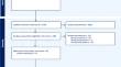
Results: From an initial search, 273 records were identified, narrowed down to 9 articles after applying our criteria. Additionally, two articles were added through citation searching. This, a total of 11 articles were incorporated in this study.
Conclusion: This micro-CT contrast agent review underscores the need for tailored choices based on research goals. Both Barium sulfate and Iodine-based agents showing excellent results, providing high resolution (micro) vascular content, especially in ex-vivo specimens. However, careful consideration of protocols and tissue characteristics remains imperative for optimizing the effectiveness of micro-CT imaging for the study of cranio-facial vascular development.
期刊介绍:
Neuroradiology aims to provide state-of-the-art medical and scientific information in the fields of Neuroradiology, Neurosciences, Neurology, Psychiatry, Neurosurgery, and related medical specialities. Neuroradiology as the official Journal of the European Society of Neuroradiology receives submissions from all parts of the world and publishes peer-reviewed original research, comprehensive reviews, educational papers, opinion papers, and short reports on exceptional clinical observations and new technical developments in the field of Neuroimaging and Neurointervention. The journal has subsections for Diagnostic and Interventional Neuroradiology, Advanced Neuroimaging, Paediatric Neuroradiology, Head-Neck-ENT Radiology, Spine Neuroradiology, and for submissions from Japan. Neuroradiology aims to provide new knowledge about and insights into the function and pathology of the human nervous system that may help to better diagnose and treat nervous system diseases. Neuroradiology is a member of the Committee on Publication Ethics (COPE) and follows the COPE core practices. Neuroradiology prefers articles that are free of bias, self-critical regarding limitations, transparent and clear in describing study participants, methods, and statistics, and short in presenting results. Before peer-review all submissions are automatically checked by iThenticate to assess for potential overlap in prior publication.


 分享
分享
 求助内容:
求助内容: 应助结果提醒方式:
 扫码关注我们
