Yuheng Liao , Chunlian Qin , Xiaoyu Zhang , Jing Ye , Zhongyuan Xu , Haotian Zong , Ning Hu , Diming Zhang
{"title":"双模式、图像增强、微型显微镜系统,用于对活细胞进行与培养箱兼容的监测。","authors":"Yuheng Liao , Chunlian Qin , Xiaoyu Zhang , Jing Ye , Zhongyuan Xu , Haotian Zong , Ning Hu , Diming Zhang","doi":"10.1016/j.talanta.2024.126537","DOIUrl":null,"url":null,"abstract":"<div><p>Imaging live cells under stable culture conditions is essential to investigate cell physiological activities and proliferation. To achieve this goal, typically, a specialized incubation chamber that creates desired culture conditions needs to be incorporated into a microscopy system to perform cell monitoring. However, such imaging systems are generally large and costly, hampering their wide applications. Recent advances in the field of miniaturized microscopy systems have enabled incubator cell monitoring, providing a hospitable environment for live cells. Although these systems are more cost-effective, they are usually limited in imaging modalities and spatial temporal resolution. Here, we present a dual-mode, image-enhanced, miniaturized microscopy system (termed MiniCube) for direct monitoring of live cells inside incubators. MiniCube enables both bright field imaging and fluorescence imaging with single-cell spatial resolution and sub-second temporal resolution. Moreover, this system can also perform cell monitoring inside the incubator with tunable time scales ranging from a few seconds to days. Meanwhile, automatic cell segmentation and image enhancement are realized by the proposed data analysis pipeline of this system, and the signal-to-noise ratio (SNR) of acquired data is significantly improved using a deep learning based image denoising algorithm. Image data can be acquired with 5 times lower light exposure while maintaining comparable SNR. The versatility of this miniaturized microscopy system lends itself to various applications in biology studies, providing a practical platform and method for studying live cell dynamics within the incubator.</p></div>","PeriodicalId":435,"journal":{"name":"Talanta","volume":"278 ","pages":"Article 126537"},"PeriodicalIF":6.1000,"publicationDate":"2024-10-01","publicationTypes":"Journal Article","fieldsOfStudy":null,"isOpenAccess":false,"openAccessPdf":"","citationCount":"0","resultStr":"{\"title\":\"A dual-mode, image-enhanced, miniaturized microscopy system for incubator-compatible monitoring of live cells\",\"authors\":\"Yuheng Liao , Chunlian Qin , Xiaoyu Zhang , Jing Ye , Zhongyuan Xu , Haotian Zong , Ning Hu , Diming Zhang\",\"doi\":\"10.1016/j.talanta.2024.126537\",\"DOIUrl\":null,\"url\":null,\"abstract\":\"<div><p>Imaging live cells under stable culture conditions is essential to investigate cell physiological activities and proliferation. To achieve this goal, typically, a specialized incubation chamber that creates desired culture conditions needs to be incorporated into a microscopy system to perform cell monitoring. However, such imaging systems are generally large and costly, hampering their wide applications. Recent advances in the field of miniaturized microscopy systems have enabled incubator cell monitoring, providing a hospitable environment for live cells. Although these systems are more cost-effective, they are usually limited in imaging modalities and spatial temporal resolution. Here, we present a dual-mode, image-enhanced, miniaturized microscopy system (termed MiniCube) for direct monitoring of live cells inside incubators. MiniCube enables both bright field imaging and fluorescence imaging with single-cell spatial resolution and sub-second temporal resolution. Moreover, this system can also perform cell monitoring inside the incubator with tunable time scales ranging from a few seconds to days. Meanwhile, automatic cell segmentation and image enhancement are realized by the proposed data analysis pipeline of this system, and the signal-to-noise ratio (SNR) of acquired data is significantly improved using a deep learning based image denoising algorithm. Image data can be acquired with 5 times lower light exposure while maintaining comparable SNR. The versatility of this miniaturized microscopy system lends itself to various applications in biology studies, providing a practical platform and method for studying live cell dynamics within the incubator.</p></div>\",\"PeriodicalId\":435,\"journal\":{\"name\":\"Talanta\",\"volume\":\"278 \",\"pages\":\"Article 126537\"},\"PeriodicalIF\":6.1000,\"publicationDate\":\"2024-10-01\",\"publicationTypes\":\"Journal Article\",\"fieldsOfStudy\":null,\"isOpenAccess\":false,\"openAccessPdf\":\"\",\"citationCount\":\"0\",\"resultStr\":null,\"platform\":\"Semanticscholar\",\"paperid\":null,\"PeriodicalName\":\"Talanta\",\"FirstCategoryId\":\"92\",\"ListUrlMain\":\"https://www.sciencedirect.com/science/article/pii/S0039914024009160\",\"RegionNum\":1,\"RegionCategory\":\"化学\",\"ArticlePicture\":[],\"TitleCN\":null,\"AbstractTextCN\":null,\"PMCID\":null,\"EPubDate\":\"2024/7/9 0:00:00\",\"PubModel\":\"Epub\",\"JCR\":\"Q1\",\"JCRName\":\"CHEMISTRY, ANALYTICAL\",\"Score\":null,\"Total\":0}","platform":"Semanticscholar","paperid":null,"PeriodicalName":"Talanta","FirstCategoryId":"92","ListUrlMain":"https://www.sciencedirect.com/science/article/pii/S0039914024009160","RegionNum":1,"RegionCategory":"化学","ArticlePicture":[],"TitleCN":null,"AbstractTextCN":null,"PMCID":null,"EPubDate":"2024/7/9 0:00:00","PubModel":"Epub","JCR":"Q1","JCRName":"CHEMISTRY, ANALYTICAL","Score":null,"Total":0}
A dual-mode, image-enhanced, miniaturized microscopy system for incubator-compatible monitoring of live cells
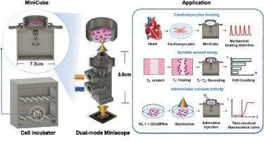
Imaging live cells under stable culture conditions is essential to investigate cell physiological activities and proliferation. To achieve this goal, typically, a specialized incubation chamber that creates desired culture conditions needs to be incorporated into a microscopy system to perform cell monitoring. However, such imaging systems are generally large and costly, hampering their wide applications. Recent advances in the field of miniaturized microscopy systems have enabled incubator cell monitoring, providing a hospitable environment for live cells. Although these systems are more cost-effective, they are usually limited in imaging modalities and spatial temporal resolution. Here, we present a dual-mode, image-enhanced, miniaturized microscopy system (termed MiniCube) for direct monitoring of live cells inside incubators. MiniCube enables both bright field imaging and fluorescence imaging with single-cell spatial resolution and sub-second temporal resolution. Moreover, this system can also perform cell monitoring inside the incubator with tunable time scales ranging from a few seconds to days. Meanwhile, automatic cell segmentation and image enhancement are realized by the proposed data analysis pipeline of this system, and the signal-to-noise ratio (SNR) of acquired data is significantly improved using a deep learning based image denoising algorithm. Image data can be acquired with 5 times lower light exposure while maintaining comparable SNR. The versatility of this miniaturized microscopy system lends itself to various applications in biology studies, providing a practical platform and method for studying live cell dynamics within the incubator.
期刊介绍:
Talanta provides a forum for the publication of original research papers, short communications, and critical reviews in all branches of pure and applied analytical chemistry. Papers are evaluated based on established guidelines, including the fundamental nature of the study, scientific novelty, substantial improvement or advantage over existing technology or methods, and demonstrated analytical applicability. Original research papers on fundamental studies, and on novel sensor and instrumentation developments, are encouraged. Novel or improved applications in areas such as clinical and biological chemistry, environmental analysis, geochemistry, materials science and engineering, and analytical platforms for omics development are welcome.
Analytical performance of methods should be determined, including interference and matrix effects, and methods should be validated by comparison with a standard method, or analysis of a certified reference material. Simple spiking recoveries may not be sufficient. The developed method should especially comprise information on selectivity, sensitivity, detection limits, accuracy, and reliability. However, applying official validation or robustness studies to a routine method or technique does not necessarily constitute novelty. Proper statistical treatment of the data should be provided. Relevant literature should be cited, including related publications by the authors, and authors should discuss how their proposed methodology compares with previously reported methods.


 分享
分享
 求助内容:
求助内容: 应助结果提醒方式:
 扫码关注我们
