A. I. Bogatyreva, E. V. Gerasimova, T. V. Kirichenko, Yu. V. Markina, T. V. Popkova, M. V. Shalygina, T. V. Tolstik, A. M. Markin, A. N. Orekhov
{"title":"免疫炎症性风湿病患者单核细胞的促炎性激活。","authors":"A. I. Bogatyreva, E. V. Gerasimova, T. V. Kirichenko, Yu. V. Markina, T. V. Popkova, M. V. Shalygina, T. V. Tolstik, A. M. Markin, A. N. Orekhov","doi":"10.1134/S1607672924700959","DOIUrl":null,"url":null,"abstract":"<p>The pathogenesis of immunoinflammatory rheumatic diseases (IRDs) is based on chronic inflammation, one of the key mechanisms of which may be abnormal activation of macrophages, leading to further disruption of the immune system.</p><p>. The objective of this study was to evaluate the proinflammatory activation of circulating monocytes in patients with IRDs.</p><p>. The study involved 149 participants (53 patients with rheumatoid arthritis (RA), 45 patients with systemic lupus erythematosus (SLE), 34 patients with systemic scleroderma (SSc), and 17 participants without IRDs) 30 to 65 years old. Basal and lipopolysaccharide (LPS)-stimulated secretion of monocytes was studied in a primary culture of monocytes obtained from blood by immunomagnetic separation. Quantitative assessment of the cytokines tumor necrosis factor α (TNF-α), interleukin 1β (IL-1β), as well as the chemokine monocyte chemoattractant protein-1 (MCP-1) was carried out in the culture fluid by ELISA. Proinflammatory activation of monocytes was calculated as the ratio of LPS-stimulated and basal secretions.</p><p>. It was shown that the basal secretion of all studied cytokines was significantly increased in all groups of patients with IRDs, except for the secretion of IL-1β in the SLE group, compared to the control. LPS-stimulated secretion of TNF-α was increased and MCP-1 was decreased in patients with IRDs compared to the control group; LPS-stimulated IL-1β secretion only in the SSc group significantly differed from the control group. In the RA group, monocyte activation was reduced for all cytokines compared to the control; in the SLE group, for TNF-α and MCP-1; in the SSc group, for MCP-1.</p><p>. The decrease in proinflammatory activation of monocytes in patients with IRDs is due to a high level of basal secretion of cytokines, which can lead to disruption of the adequate immune response in these diseases and is an important link in the pathogenesis of chronic inflammation.</p>","PeriodicalId":529,"journal":{"name":"Doklady Biochemistry and Biophysics","volume":"517 1","pages":"228 - 234"},"PeriodicalIF":0.8000,"publicationDate":"2024-07-13","publicationTypes":"Journal Article","fieldsOfStudy":null,"isOpenAccess":false,"openAccessPdf":"","citationCount":"0","resultStr":"{\"title\":\"Proinflammatory Activation of Monocytes in Patients with Immunoinflammatory Rheumatic Diseases\",\"authors\":\"A. I. Bogatyreva, E. V. Gerasimova, T. V. Kirichenko, Yu. V. Markina, T. V. Popkova, M. V. Shalygina, T. V. Tolstik, A. M. Markin, A. N. Orekhov\",\"doi\":\"10.1134/S1607672924700959\",\"DOIUrl\":null,\"url\":null,\"abstract\":\"<p>The pathogenesis of immunoinflammatory rheumatic diseases (IRDs) is based on chronic inflammation, one of the key mechanisms of which may be abnormal activation of macrophages, leading to further disruption of the immune system.</p><p>. The objective of this study was to evaluate the proinflammatory activation of circulating monocytes in patients with IRDs.</p><p>. The study involved 149 participants (53 patients with rheumatoid arthritis (RA), 45 patients with systemic lupus erythematosus (SLE), 34 patients with systemic scleroderma (SSc), and 17 participants without IRDs) 30 to 65 years old. Basal and lipopolysaccharide (LPS)-stimulated secretion of monocytes was studied in a primary culture of monocytes obtained from blood by immunomagnetic separation. Quantitative assessment of the cytokines tumor necrosis factor α (TNF-α), interleukin 1β (IL-1β), as well as the chemokine monocyte chemoattractant protein-1 (MCP-1) was carried out in the culture fluid by ELISA. Proinflammatory activation of monocytes was calculated as the ratio of LPS-stimulated and basal secretions.</p><p>. It was shown that the basal secretion of all studied cytokines was significantly increased in all groups of patients with IRDs, except for the secretion of IL-1β in the SLE group, compared to the control. LPS-stimulated secretion of TNF-α was increased and MCP-1 was decreased in patients with IRDs compared to the control group; LPS-stimulated IL-1β secretion only in the SSc group significantly differed from the control group. In the RA group, monocyte activation was reduced for all cytokines compared to the control; in the SLE group, for TNF-α and MCP-1; in the SSc group, for MCP-1.</p><p>. The decrease in proinflammatory activation of monocytes in patients with IRDs is due to a high level of basal secretion of cytokines, which can lead to disruption of the adequate immune response in these diseases and is an important link in the pathogenesis of chronic inflammation.</p>\",\"PeriodicalId\":529,\"journal\":{\"name\":\"Doklady Biochemistry and Biophysics\",\"volume\":\"517 1\",\"pages\":\"228 - 234\"},\"PeriodicalIF\":0.8000,\"publicationDate\":\"2024-07-13\",\"publicationTypes\":\"Journal Article\",\"fieldsOfStudy\":null,\"isOpenAccess\":false,\"openAccessPdf\":\"\",\"citationCount\":\"0\",\"resultStr\":null,\"platform\":\"Semanticscholar\",\"paperid\":null,\"PeriodicalName\":\"Doklady Biochemistry and Biophysics\",\"FirstCategoryId\":\"99\",\"ListUrlMain\":\"https://link.springer.com/article/10.1134/S1607672924700959\",\"RegionNum\":4,\"RegionCategory\":\"生物学\",\"ArticlePicture\":[],\"TitleCN\":null,\"AbstractTextCN\":null,\"PMCID\":null,\"EPubDate\":\"\",\"PubModel\":\"\",\"JCR\":\"Q4\",\"JCRName\":\"BIOCHEMISTRY & MOLECULAR BIOLOGY\",\"Score\":null,\"Total\":0}","platform":"Semanticscholar","paperid":null,"PeriodicalName":"Doklady Biochemistry and Biophysics","FirstCategoryId":"99","ListUrlMain":"https://link.springer.com/article/10.1134/S1607672924700959","RegionNum":4,"RegionCategory":"生物学","ArticlePicture":[],"TitleCN":null,"AbstractTextCN":null,"PMCID":null,"EPubDate":"","PubModel":"","JCR":"Q4","JCRName":"BIOCHEMISTRY & MOLECULAR BIOLOGY","Score":null,"Total":0}
Proinflammatory Activation of Monocytes in Patients with Immunoinflammatory Rheumatic Diseases
The pathogenesis of immunoinflammatory rheumatic diseases (IRDs) is based on chronic inflammation, one of the key mechanisms of which may be abnormal activation of macrophages, leading to further disruption of the immune system.
. The objective of this study was to evaluate the proinflammatory activation of circulating monocytes in patients with IRDs.
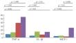
. The study involved 149 participants (53 patients with rheumatoid arthritis (RA), 45 patients with systemic lupus erythematosus (SLE), 34 patients with systemic scleroderma (SSc), and 17 participants without IRDs) 30 to 65 years old. Basal and lipopolysaccharide (LPS)-stimulated secretion of monocytes was studied in a primary culture of monocytes obtained from blood by immunomagnetic separation. Quantitative assessment of the cytokines tumor necrosis factor α (TNF-α), interleukin 1β (IL-1β), as well as the chemokine monocyte chemoattractant protein-1 (MCP-1) was carried out in the culture fluid by ELISA. Proinflammatory activation of monocytes was calculated as the ratio of LPS-stimulated and basal secretions.
. It was shown that the basal secretion of all studied cytokines was significantly increased in all groups of patients with IRDs, except for the secretion of IL-1β in the SLE group, compared to the control. LPS-stimulated secretion of TNF-α was increased and MCP-1 was decreased in patients with IRDs compared to the control group; LPS-stimulated IL-1β secretion only in the SSc group significantly differed from the control group. In the RA group, monocyte activation was reduced for all cytokines compared to the control; in the SLE group, for TNF-α and MCP-1; in the SSc group, for MCP-1.
. The decrease in proinflammatory activation of monocytes in patients with IRDs is due to a high level of basal secretion of cytokines, which can lead to disruption of the adequate immune response in these diseases and is an important link in the pathogenesis of chronic inflammation.
期刊介绍:
Doklady Biochemistry and Biophysics is a journal consisting of English translations of articles published in Russian in biochemistry and biophysics sections of the Russian-language journal Doklady Akademii Nauk. The journal''s goal is to publish the most significant new research in biochemistry and biophysics carried out in Russia today or in collaboration with Russian authors. The journal accepts only articles in the Russian language that are submitted or recommended by acting Russian or foreign members of the Russian Academy of Sciences. The journal does not accept direct submissions in English.


 分享
分享
 求助内容:
求助内容: 应助结果提醒方式:
 扫码关注我们
