{"title":"重新评估股外侧周动脉解剖结构的多样性及其临床应用性:尸体研究。","authors":"Anju Chaudhary, Apurba Patra, Pooja Garg","doi":"10.5115/acb.24.047","DOIUrl":null,"url":null,"abstract":"<p><p>Studies reveal variations in the in the origin, number, and branching patterns of the lateral circumflex femoral artery (LCFA). The present study aimed to document such variations and their potential clinical applicability. Thirty-two femoral triangles of 16 embalmed adult human cadavers were dissected to investigate the variation in the origin, number, and branching patterns of LCFA. The main branches of the LCFA were tracked independently for numerical variations in branching pattern. The distance between the origin of LCFA and mid inguinal point (MIP) was also measured in each case. LCFA was most commonly arising from profunda femoris (PF), followed by femoral artery (FA) and common trunk of the femoral artery (CFA). Duplication LCFA was observed in 15 (46.87%) limbs, in 5 (31.25%) cases duplication was only on right side, in 4 (25%) cases duplication was only on left side and in 3 (18.75%), duplication was bilateral. Cases with duplication of LCFA, showed numerical variations with descending pattern being the most common. The average distance of LCFA1 and LCFA2 from mid-inguinal point was 5.77±1.35 cm and 6.14±2.05 cm respectively. Detailed information regarding the occurrence of duplication will be great importance for surgeons, interventional radiologists, and other medical professionals performing procedures in the femoral region. Knowledge of variation of branching pattern of LCFA is utmost important as surgeons use the descending branch of the LCFA in bypass grafting and vascular reconstruction surgeries.</p>","PeriodicalId":7831,"journal":{"name":"Anatomy & Cell Biology","volume":" ","pages":"346-352"},"PeriodicalIF":1.1000,"publicationDate":"2024-09-30","publicationTypes":"Journal Article","fieldsOfStudy":null,"isOpenAccess":false,"openAccessPdf":"https://www.ncbi.nlm.nih.gov/pmc/articles/PMC11424569/pdf/","citationCount":"0","resultStr":"{\"title\":\"Reappraisal of anatomical diversity of lateral circumflex femoral artery with its substantial clinical applicability: cadaveric study.\",\"authors\":\"Anju Chaudhary, Apurba Patra, Pooja Garg\",\"doi\":\"10.5115/acb.24.047\",\"DOIUrl\":null,\"url\":null,\"abstract\":\"<p><p>Studies reveal variations in the in the origin, number, and branching patterns of the lateral circumflex femoral artery (LCFA). The present study aimed to document such variations and their potential clinical applicability. Thirty-two femoral triangles of 16 embalmed adult human cadavers were dissected to investigate the variation in the origin, number, and branching patterns of LCFA. The main branches of the LCFA were tracked independently for numerical variations in branching pattern. The distance between the origin of LCFA and mid inguinal point (MIP) was also measured in each case. LCFA was most commonly arising from profunda femoris (PF), followed by femoral artery (FA) and common trunk of the femoral artery (CFA). Duplication LCFA was observed in 15 (46.87%) limbs, in 5 (31.25%) cases duplication was only on right side, in 4 (25%) cases duplication was only on left side and in 3 (18.75%), duplication was bilateral. Cases with duplication of LCFA, showed numerical variations with descending pattern being the most common. The average distance of LCFA1 and LCFA2 from mid-inguinal point was 5.77±1.35 cm and 6.14±2.05 cm respectively. Detailed information regarding the occurrence of duplication will be great importance for surgeons, interventional radiologists, and other medical professionals performing procedures in the femoral region. Knowledge of variation of branching pattern of LCFA is utmost important as surgeons use the descending branch of the LCFA in bypass grafting and vascular reconstruction surgeries.</p>\",\"PeriodicalId\":7831,\"journal\":{\"name\":\"Anatomy & Cell Biology\",\"volume\":\" \",\"pages\":\"346-352\"},\"PeriodicalIF\":1.1000,\"publicationDate\":\"2024-09-30\",\"publicationTypes\":\"Journal Article\",\"fieldsOfStudy\":null,\"isOpenAccess\":false,\"openAccessPdf\":\"https://www.ncbi.nlm.nih.gov/pmc/articles/PMC11424569/pdf/\",\"citationCount\":\"0\",\"resultStr\":null,\"platform\":\"Semanticscholar\",\"paperid\":null,\"PeriodicalName\":\"Anatomy & Cell Biology\",\"FirstCategoryId\":\"1085\",\"ListUrlMain\":\"https://doi.org/10.5115/acb.24.047\",\"RegionNum\":0,\"RegionCategory\":null,\"ArticlePicture\":[],\"TitleCN\":null,\"AbstractTextCN\":null,\"PMCID\":null,\"EPubDate\":\"2024/7/15 0:00:00\",\"PubModel\":\"Epub\",\"JCR\":\"Q3\",\"JCRName\":\"ANATOMY & MORPHOLOGY\",\"Score\":null,\"Total\":0}","platform":"Semanticscholar","paperid":null,"PeriodicalName":"Anatomy & Cell Biology","FirstCategoryId":"1085","ListUrlMain":"https://doi.org/10.5115/acb.24.047","RegionNum":0,"RegionCategory":null,"ArticlePicture":[],"TitleCN":null,"AbstractTextCN":null,"PMCID":null,"EPubDate":"2024/7/15 0:00:00","PubModel":"Epub","JCR":"Q3","JCRName":"ANATOMY & MORPHOLOGY","Score":null,"Total":0}
Reappraisal of anatomical diversity of lateral circumflex femoral artery with its substantial clinical applicability: cadaveric study.
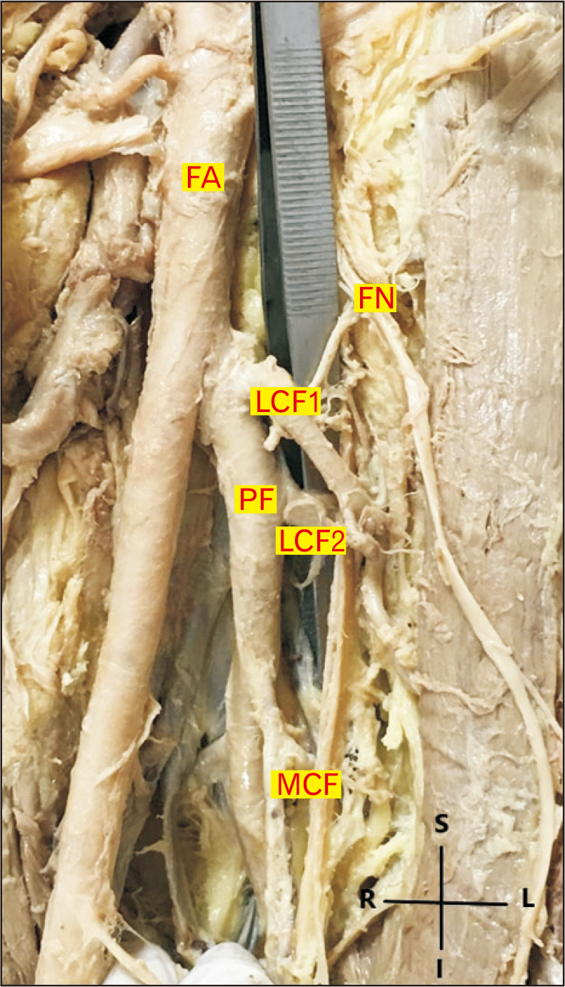
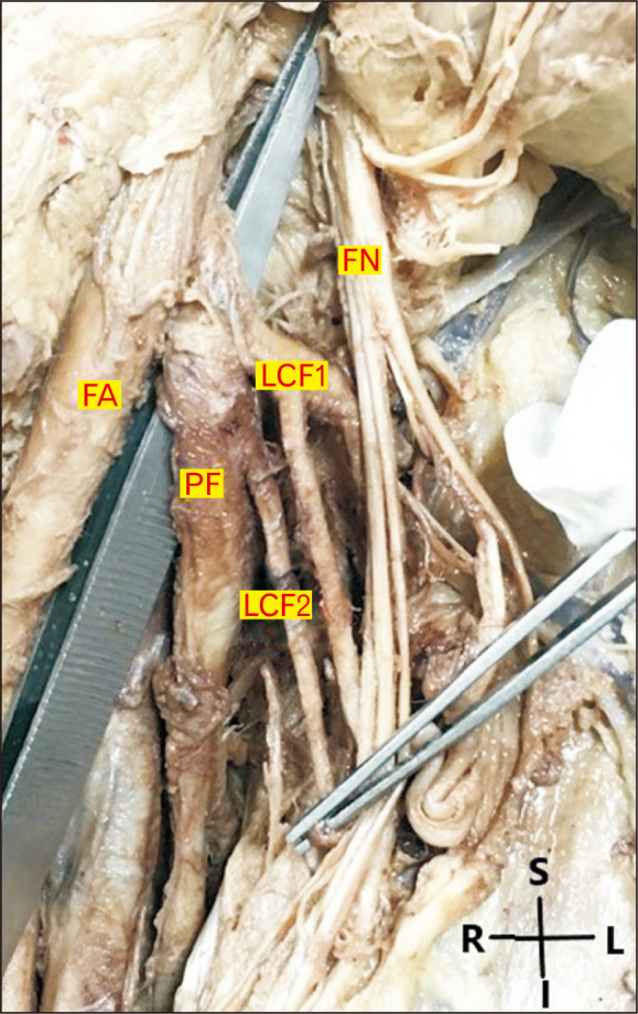
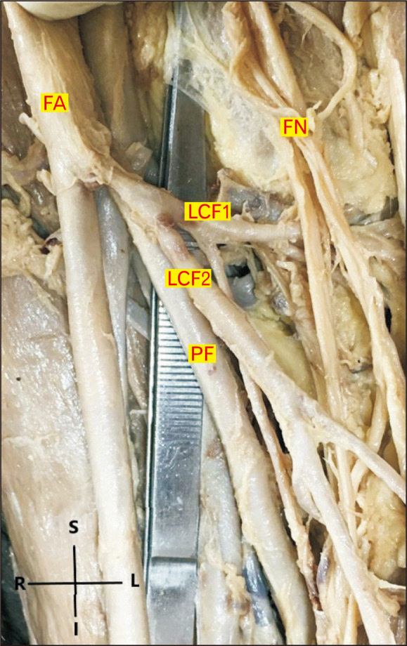
Studies reveal variations in the in the origin, number, and branching patterns of the lateral circumflex femoral artery (LCFA). The present study aimed to document such variations and their potential clinical applicability. Thirty-two femoral triangles of 16 embalmed adult human cadavers were dissected to investigate the variation in the origin, number, and branching patterns of LCFA. The main branches of the LCFA were tracked independently for numerical variations in branching pattern. The distance between the origin of LCFA and mid inguinal point (MIP) was also measured in each case. LCFA was most commonly arising from profunda femoris (PF), followed by femoral artery (FA) and common trunk of the femoral artery (CFA). Duplication LCFA was observed in 15 (46.87%) limbs, in 5 (31.25%) cases duplication was only on right side, in 4 (25%) cases duplication was only on left side and in 3 (18.75%), duplication was bilateral. Cases with duplication of LCFA, showed numerical variations with descending pattern being the most common. The average distance of LCFA1 and LCFA2 from mid-inguinal point was 5.77±1.35 cm and 6.14±2.05 cm respectively. Detailed information regarding the occurrence of duplication will be great importance for surgeons, interventional radiologists, and other medical professionals performing procedures in the femoral region. Knowledge of variation of branching pattern of LCFA is utmost important as surgeons use the descending branch of the LCFA in bypass grafting and vascular reconstruction surgeries.


 分享
分享
 求助内容:
求助内容: 应助结果提醒方式:
 扫码关注我们
