{"title":"一例从 Descemet's Stripping Automated Endothelial Keratoplasty 移除脱位移植物后仍能保持透明角膜的病例。","authors":"Yuta Nariya, Takashi Ono, Yuichi Asahina, Atsushi Kondo, Yukako Taketani, Mikiko Kimakura, Tetsuya Toyono, Makoto Aihara, Takashi Miyai","doi":"10.1159/000539392","DOIUrl":null,"url":null,"abstract":"<p><strong>Introduction: </strong>Corneal graft detachment is a major postoperative complication of Descemet's stripping automated endothelial keratoplasty (DSAEK). When a corneal graft becomes detached, corneal endothelial function generally fails, and repeat corneal transplantation is required. Herein, we report a rare case in which a transparent cornea was maintained after the removal of a dislocated DSAEK graft.</p><p><strong>Case presentation: </strong>A 79-year-old woman with a residual lens cortex who had undergone cataract surgery was referred to our hospital. The cortex was removed, and bullous keratopathy progressed. Six months after the initial surgery, DSAEK was performed under topical anesthesia without any complications. Although the corneal graft had attached fairly well, it detached from the host cornea 3 weeks later. Two months after DSAEK, an air tamponade was used to treat the anterior chamber with single interrupted suturing; however, the graft detached again, except for the suture site. Because the detached cornea became cloudy in the anterior chamber, it was surgically removed 8 months after DSAEK. Accordingly, the host cornea transparency improved to a best-corrected visual acuity of 0.8 with a rigid gas permeable lens and a central corneal thickness of 580 μm. The corneal endothelial cell density was 995 cells/mm<sup>2</sup>.</p><p><strong>Conclusion: </strong>Removal of the corneal graft from the dislocated cloudy graft improved the visual acuity of this patient after DSAEK. The condition of the cornea should be carefully monitored after corneal endothelial transplantation, even after the graft has been dislocated.</p>","PeriodicalId":9635,"journal":{"name":"Case Reports in Ophthalmology","volume":"15 1","pages":"518-524"},"PeriodicalIF":0.6000,"publicationDate":"2024-06-26","publicationTypes":"Journal Article","fieldsOfStudy":null,"isOpenAccess":false,"openAccessPdf":"https://www.ncbi.nlm.nih.gov/pmc/articles/PMC11250565/pdf/","citationCount":"0","resultStr":"{\"title\":\"A Case of Transparent Cornea Maintained after Removal of Dislocated Graft from Descemet's Stripping Automated Endothelial Keratoplasty.\",\"authors\":\"Yuta Nariya, Takashi Ono, Yuichi Asahina, Atsushi Kondo, Yukako Taketani, Mikiko Kimakura, Tetsuya Toyono, Makoto Aihara, Takashi Miyai\",\"doi\":\"10.1159/000539392\",\"DOIUrl\":null,\"url\":null,\"abstract\":\"<p><strong>Introduction: </strong>Corneal graft detachment is a major postoperative complication of Descemet's stripping automated endothelial keratoplasty (DSAEK). When a corneal graft becomes detached, corneal endothelial function generally fails, and repeat corneal transplantation is required. Herein, we report a rare case in which a transparent cornea was maintained after the removal of a dislocated DSAEK graft.</p><p><strong>Case presentation: </strong>A 79-year-old woman with a residual lens cortex who had undergone cataract surgery was referred to our hospital. The cortex was removed, and bullous keratopathy progressed. Six months after the initial surgery, DSAEK was performed under topical anesthesia without any complications. Although the corneal graft had attached fairly well, it detached from the host cornea 3 weeks later. Two months after DSAEK, an air tamponade was used to treat the anterior chamber with single interrupted suturing; however, the graft detached again, except for the suture site. Because the detached cornea became cloudy in the anterior chamber, it was surgically removed 8 months after DSAEK. Accordingly, the host cornea transparency improved to a best-corrected visual acuity of 0.8 with a rigid gas permeable lens and a central corneal thickness of 580 μm. The corneal endothelial cell density was 995 cells/mm<sup>2</sup>.</p><p><strong>Conclusion: </strong>Removal of the corneal graft from the dislocated cloudy graft improved the visual acuity of this patient after DSAEK. The condition of the cornea should be carefully monitored after corneal endothelial transplantation, even after the graft has been dislocated.</p>\",\"PeriodicalId\":9635,\"journal\":{\"name\":\"Case Reports in Ophthalmology\",\"volume\":\"15 1\",\"pages\":\"518-524\"},\"PeriodicalIF\":0.6000,\"publicationDate\":\"2024-06-26\",\"publicationTypes\":\"Journal Article\",\"fieldsOfStudy\":null,\"isOpenAccess\":false,\"openAccessPdf\":\"https://www.ncbi.nlm.nih.gov/pmc/articles/PMC11250565/pdf/\",\"citationCount\":\"0\",\"resultStr\":null,\"platform\":\"Semanticscholar\",\"paperid\":null,\"PeriodicalName\":\"Case Reports in Ophthalmology\",\"FirstCategoryId\":\"1085\",\"ListUrlMain\":\"https://doi.org/10.1159/000539392\",\"RegionNum\":0,\"RegionCategory\":null,\"ArticlePicture\":[],\"TitleCN\":null,\"AbstractTextCN\":null,\"PMCID\":null,\"EPubDate\":\"2024/1/1 0:00:00\",\"PubModel\":\"eCollection\",\"JCR\":\"Q4\",\"JCRName\":\"OPHTHALMOLOGY\",\"Score\":null,\"Total\":0}","platform":"Semanticscholar","paperid":null,"PeriodicalName":"Case Reports in Ophthalmology","FirstCategoryId":"1085","ListUrlMain":"https://doi.org/10.1159/000539392","RegionNum":0,"RegionCategory":null,"ArticlePicture":[],"TitleCN":null,"AbstractTextCN":null,"PMCID":null,"EPubDate":"2024/1/1 0:00:00","PubModel":"eCollection","JCR":"Q4","JCRName":"OPHTHALMOLOGY","Score":null,"Total":0}
A Case of Transparent Cornea Maintained after Removal of Dislocated Graft from Descemet's Stripping Automated Endothelial Keratoplasty.
Introduction: Corneal graft detachment is a major postoperative complication of Descemet's stripping automated endothelial keratoplasty (DSAEK). When a corneal graft becomes detached, corneal endothelial function generally fails, and repeat corneal transplantation is required. Herein, we report a rare case in which a transparent cornea was maintained after the removal of a dislocated DSAEK graft.
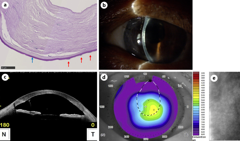
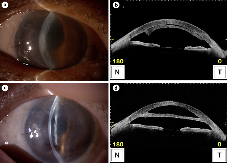
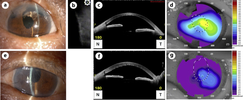
Case presentation: A 79-year-old woman with a residual lens cortex who had undergone cataract surgery was referred to our hospital. The cortex was removed, and bullous keratopathy progressed. Six months after the initial surgery, DSAEK was performed under topical anesthesia without any complications. Although the corneal graft had attached fairly well, it detached from the host cornea 3 weeks later. Two months after DSAEK, an air tamponade was used to treat the anterior chamber with single interrupted suturing; however, the graft detached again, except for the suture site. Because the detached cornea became cloudy in the anterior chamber, it was surgically removed 8 months after DSAEK. Accordingly, the host cornea transparency improved to a best-corrected visual acuity of 0.8 with a rigid gas permeable lens and a central corneal thickness of 580 μm. The corneal endothelial cell density was 995 cells/mm2.
Conclusion: Removal of the corneal graft from the dislocated cloudy graft improved the visual acuity of this patient after DSAEK. The condition of the cornea should be carefully monitored after corneal endothelial transplantation, even after the graft has been dislocated.
期刊介绍:
This peer-reviewed online-only journal publishes original case reports covering the entire spectrum of ophthalmology, including prevention, diagnosis, treatment, toxicities of therapy, supportive care, quality-of-life, and survivorship issues. The submission of negative results is strongly encouraged. The journal will also accept case reports dealing with the use of novel technologies, both in the arena of diagnosis and treatment. Supplementary material is welcomed. The intent of the journal is to provide clinicians and researchers with a tool to disseminate their personal experiences to a wider public as well as to review interesting cases encountered by colleagues all over the world. Universally used terms can be searched across the entire growing collection of case reports, further facilitating the retrieval of specific information. Following the open access principle, the entire contents can be retrieved at no charge, guaranteeing easy access to this valuable source of anecdotal information at all times.


 分享
分享
 求助内容:
求助内容: 应助结果提醒方式:
 扫码关注我们
