Frederico Portugal-Gaspar, Antonio Lopez-Beltran, Gladell P Paner, Ana Blanca, Enrique Gómez Gómez, Rodolfo Montironi, Alessia Cimadamore, Andreia Bilé, Metka Volavšek, Liang Cheng
{"title":"膀胱巨细胞癌 :临床病理分析和肿瘤学结果。","authors":"Frederico Portugal-Gaspar, Antonio Lopez-Beltran, Gladell P Paner, Ana Blanca, Enrique Gómez Gómez, Rodolfo Montironi, Alessia Cimadamore, Andreia Bilé, Metka Volavšek, Liang Cheng","doi":"10.1007/s00428-024-03858-w","DOIUrl":null,"url":null,"abstract":"<p><p>We present the clinicopathological features of 23 cases of the giant cell subtype of urothelial carcinoma, a rare subtype of bladder cancer recognized in the current World Health Organization classification of urological tumors. Histologically, the architectural pattern of the tumor varied from infiltrating to the solid expansile pleomorphic tumor with giant, bizarre, anaplastic cells. Typical or atypical mitotic figures were frequently present in all cases. Between 10 and 30% of the tumor had a giant cell component. All cases were associated with conventional high-grade urothelial carcinoma, with areas of squamous cell divergent differentiation and micropapillary carcinoma present in six and two cases, respectively. In one case each had sarcomatoid, nested, small cell, or glandular divergent differentiation. At diagnosis, 35% of patients had advanced disease and 12% had distant metastases. When comparing giant cell urothelial carcinoma with conventional urothelial carcinoma in a matched analysis, differences in overall and cancer-specific survival were observed, particularly in the T1 stage category. Immunohistochemical staining showed a similar profile of urothelial lineage with frequent positive expression of uroplakin II, GATA3, CK20, CK7, and S100P in both giant cell and conventional urothelial carcinomas. High Ki67 proliferation (range, 60-90%; mean, 71%) and nuclear p53 accumulation (mutant profile; range, 50-90%; mean, 64%) were observed. Using the 22C3 assay, the expression of PD-L1 was found to be variable in two cases, and beta-HCG was negative. In conclusion, giant cell carcinoma is a subtype of urothelial carcinoma associated with advanced clinical stage and a trend to lower survival rates.</p>","PeriodicalId":23514,"journal":{"name":"Virchows Archiv","volume":" ","pages":"535-546"},"PeriodicalIF":3.1000,"publicationDate":"2024-09-01","publicationTypes":"Journal Article","fieldsOfStudy":null,"isOpenAccess":false,"openAccessPdf":"https://www.ncbi.nlm.nih.gov/pmc/articles/PMC11415457/pdf/","citationCount":"0","resultStr":"{\"title\":\"Giant cell carcinoma of the urinary bladder : Clinicopathologic analysis and oncological outcomes.\",\"authors\":\"Frederico Portugal-Gaspar, Antonio Lopez-Beltran, Gladell P Paner, Ana Blanca, Enrique Gómez Gómez, Rodolfo Montironi, Alessia Cimadamore, Andreia Bilé, Metka Volavšek, Liang Cheng\",\"doi\":\"10.1007/s00428-024-03858-w\",\"DOIUrl\":null,\"url\":null,\"abstract\":\"<p><p>We present the clinicopathological features of 23 cases of the giant cell subtype of urothelial carcinoma, a rare subtype of bladder cancer recognized in the current World Health Organization classification of urological tumors. Histologically, the architectural pattern of the tumor varied from infiltrating to the solid expansile pleomorphic tumor with giant, bizarre, anaplastic cells. Typical or atypical mitotic figures were frequently present in all cases. Between 10 and 30% of the tumor had a giant cell component. All cases were associated with conventional high-grade urothelial carcinoma, with areas of squamous cell divergent differentiation and micropapillary carcinoma present in six and two cases, respectively. In one case each had sarcomatoid, nested, small cell, or glandular divergent differentiation. At diagnosis, 35% of patients had advanced disease and 12% had distant metastases. When comparing giant cell urothelial carcinoma with conventional urothelial carcinoma in a matched analysis, differences in overall and cancer-specific survival were observed, particularly in the T1 stage category. Immunohistochemical staining showed a similar profile of urothelial lineage with frequent positive expression of uroplakin II, GATA3, CK20, CK7, and S100P in both giant cell and conventional urothelial carcinomas. High Ki67 proliferation (range, 60-90%; mean, 71%) and nuclear p53 accumulation (mutant profile; range, 50-90%; mean, 64%) were observed. Using the 22C3 assay, the expression of PD-L1 was found to be variable in two cases, and beta-HCG was negative. In conclusion, giant cell carcinoma is a subtype of urothelial carcinoma associated with advanced clinical stage and a trend to lower survival rates.</p>\",\"PeriodicalId\":23514,\"journal\":{\"name\":\"Virchows Archiv\",\"volume\":\" \",\"pages\":\"535-546\"},\"PeriodicalIF\":3.1000,\"publicationDate\":\"2024-09-01\",\"publicationTypes\":\"Journal Article\",\"fieldsOfStudy\":null,\"isOpenAccess\":false,\"openAccessPdf\":\"https://www.ncbi.nlm.nih.gov/pmc/articles/PMC11415457/pdf/\",\"citationCount\":\"0\",\"resultStr\":null,\"platform\":\"Semanticscholar\",\"paperid\":null,\"PeriodicalName\":\"Virchows Archiv\",\"FirstCategoryId\":\"3\",\"ListUrlMain\":\"https://doi.org/10.1007/s00428-024-03858-w\",\"RegionNum\":3,\"RegionCategory\":\"医学\",\"ArticlePicture\":[],\"TitleCN\":null,\"AbstractTextCN\":null,\"PMCID\":null,\"EPubDate\":\"2024/7/18 0:00:00\",\"PubModel\":\"Epub\",\"JCR\":\"Q1\",\"JCRName\":\"PATHOLOGY\",\"Score\":null,\"Total\":0}","platform":"Semanticscholar","paperid":null,"PeriodicalName":"Virchows Archiv","FirstCategoryId":"3","ListUrlMain":"https://doi.org/10.1007/s00428-024-03858-w","RegionNum":3,"RegionCategory":"医学","ArticlePicture":[],"TitleCN":null,"AbstractTextCN":null,"PMCID":null,"EPubDate":"2024/7/18 0:00:00","PubModel":"Epub","JCR":"Q1","JCRName":"PATHOLOGY","Score":null,"Total":0}
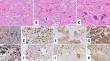
Giant cell carcinoma of the urinary bladder : Clinicopathologic analysis and oncological outcomes.
We present the clinicopathological features of 23 cases of the giant cell subtype of urothelial carcinoma, a rare subtype of bladder cancer recognized in the current World Health Organization classification of urological tumors. Histologically, the architectural pattern of the tumor varied from infiltrating to the solid expansile pleomorphic tumor with giant, bizarre, anaplastic cells. Typical or atypical mitotic figures were frequently present in all cases. Between 10 and 30% of the tumor had a giant cell component. All cases were associated with conventional high-grade urothelial carcinoma, with areas of squamous cell divergent differentiation and micropapillary carcinoma present in six and two cases, respectively. In one case each had sarcomatoid, nested, small cell, or glandular divergent differentiation. At diagnosis, 35% of patients had advanced disease and 12% had distant metastases. When comparing giant cell urothelial carcinoma with conventional urothelial carcinoma in a matched analysis, differences in overall and cancer-specific survival were observed, particularly in the T1 stage category. Immunohistochemical staining showed a similar profile of urothelial lineage with frequent positive expression of uroplakin II, GATA3, CK20, CK7, and S100P in both giant cell and conventional urothelial carcinomas. High Ki67 proliferation (range, 60-90%; mean, 71%) and nuclear p53 accumulation (mutant profile; range, 50-90%; mean, 64%) were observed. Using the 22C3 assay, the expression of PD-L1 was found to be variable in two cases, and beta-HCG was negative. In conclusion, giant cell carcinoma is a subtype of urothelial carcinoma associated with advanced clinical stage and a trend to lower survival rates.
期刊介绍:
Manuscripts of original studies reinforcing the evidence base of modern diagnostic pathology, using immunocytochemical, molecular and ultrastructural techniques, will be welcomed. In addition, papers on critical evaluation of diagnostic criteria but also broadsheets and guidelines with a solid evidence base will be considered. Consideration will also be given to reports of work in other fields relevant to the understanding of human pathology as well as manuscripts on the application of new methods and techniques in pathology. Submission of purely experimental articles is discouraged but manuscripts on experimental work applicable to diagnostic pathology are welcomed. Biomarker studies are welcomed but need to abide by strict rules (e.g. REMARK) of adequate sample size and relevant marker choice. Single marker studies on limited patient series without validated application will as a rule not be considered. Case reports will only be considered when they provide substantial new information with an impact on understanding disease or diagnostic practice.


 分享
分享
 求助内容:
求助内容: 应助结果提醒方式:
 扫码关注我们
