Bharat Rekhi, Josephine K Dermawan, Karen J Fritchie, Annette Zimpfer, Tareq M Mohammad, Fatima S Ali, Koushik Nandy, Youran Zou, Robert Stoehr, Abbas Agaimy
{"title":"EWSR1::ATF1融合是一组腹腔外上皮样和圆形细胞间充质肿瘤的特征,在表型上与硬化性上皮样纤维肉瘤和腹腔内FET::CREB融合肿瘤重叠。","authors":"Bharat Rekhi, Josephine K Dermawan, Karen J Fritchie, Annette Zimpfer, Tareq M Mohammad, Fatima S Ali, Koushik Nandy, Youran Zou, Robert Stoehr, Abbas Agaimy","doi":"10.1007/s00428-024-03879-5","DOIUrl":null,"url":null,"abstract":"<p><p>With the increasing use of next generation sequencing in soft tissue pathology, particularly in neoplasms not fitting any World Health Organization (WHO) category, the spectrum of EWSR1 fusion-associated soft tissue neoplasms has been expanding significantly. Although recurrent EWSR1::ATF1 fusions were initially limited to a triad of mesenchymal neoplasms including clear cell sarcoma of soft tissue, angiomatoid fibrous histiocytoma and malignant gastrointestinal neuroectodermal tumor (MGNET), this family has been expanding. We herein describe 4 unclassified extra-abdominal soft tissue (n = 3) and bone (n = 1) neoplasms displaying epithelioid and round cell morphology and carrying an EWSR1::ATF1 fusion. Affected were 3 males and 1 female aged 20-56 years. All primary tumors were extra-abdominal and deep-seated (chest wall, mediastinum, deltoid, and parapharyngeal soft tissue). Their size ranged 4.4-7.5 cm (median, 6.2). One patient presented with constitutional symptoms. Surgery with (2) or without (1) neo/adjuvant therapy was the treatment. At last follow-up (8-21 months), 2 patients developed progressive disease (1 recurrence; 1 distant metastasis). The immunophenotype of these tumors is potentially misleading with variable expression of EMA (2 of 3), pankeratin (2 of 4), synaptophysin (2 of 3), MUC4 (1 of 3), and ALK (1 of 3). All tumors were negative for S100 and SOX10. These observations point to the existence of heretofore under-recognized group of epithelioid and round cell neoplasms of soft tissue and bone, driven by EWSR1::ATF1 fusions, but distinct from established EWSR1::ATF1-associated soft tissue entities. Their overall morphology and immunophenotype recapitulate that of the emerging EWSR1/FUS::CREB fusion associated intra-abdominal epithelioid/round cell neoplasms. Our cases point to a potentially aggressive clinical behavior. Recognizing this tumor type is mandatory to delineate any inherent biological and/or therapeutic distinctness from other, better-known sarcomas in the differential diagnosis including sclerosing epithelioid fibrosarcoma.</p>","PeriodicalId":23514,"journal":{"name":"Virchows Archiv","volume":" ","pages":"995-1005"},"PeriodicalIF":3.0000,"publicationDate":"2024-12-01","publicationTypes":"Journal Article","fieldsOfStudy":null,"isOpenAccess":false,"openAccessPdf":"https://www.ncbi.nlm.nih.gov/pmc/articles/PMC11666693/pdf/","citationCount":"0","resultStr":"{\"title\":\"EWSR1::ATF1 fusions characterize a group of extra-abdominal epithelioid and round cell mesenchymal neoplasms, phenotypically overlapping with sclerosing epithelioid fibrosarcomas, and intra-abdominal FET::CREB fusion neoplasms.\",\"authors\":\"Bharat Rekhi, Josephine K Dermawan, Karen J Fritchie, Annette Zimpfer, Tareq M Mohammad, Fatima S Ali, Koushik Nandy, Youran Zou, Robert Stoehr, Abbas Agaimy\",\"doi\":\"10.1007/s00428-024-03879-5\",\"DOIUrl\":null,\"url\":null,\"abstract\":\"<p><p>With the increasing use of next generation sequencing in soft tissue pathology, particularly in neoplasms not fitting any World Health Organization (WHO) category, the spectrum of EWSR1 fusion-associated soft tissue neoplasms has been expanding significantly. Although recurrent EWSR1::ATF1 fusions were initially limited to a triad of mesenchymal neoplasms including clear cell sarcoma of soft tissue, angiomatoid fibrous histiocytoma and malignant gastrointestinal neuroectodermal tumor (MGNET), this family has been expanding. We herein describe 4 unclassified extra-abdominal soft tissue (n = 3) and bone (n = 1) neoplasms displaying epithelioid and round cell morphology and carrying an EWSR1::ATF1 fusion. Affected were 3 males and 1 female aged 20-56 years. All primary tumors were extra-abdominal and deep-seated (chest wall, mediastinum, deltoid, and parapharyngeal soft tissue). Their size ranged 4.4-7.5 cm (median, 6.2). One patient presented with constitutional symptoms. Surgery with (2) or without (1) neo/adjuvant therapy was the treatment. At last follow-up (8-21 months), 2 patients developed progressive disease (1 recurrence; 1 distant metastasis). The immunophenotype of these tumors is potentially misleading with variable expression of EMA (2 of 3), pankeratin (2 of 4), synaptophysin (2 of 3), MUC4 (1 of 3), and ALK (1 of 3). All tumors were negative for S100 and SOX10. These observations point to the existence of heretofore under-recognized group of epithelioid and round cell neoplasms of soft tissue and bone, driven by EWSR1::ATF1 fusions, but distinct from established EWSR1::ATF1-associated soft tissue entities. Their overall morphology and immunophenotype recapitulate that of the emerging EWSR1/FUS::CREB fusion associated intra-abdominal epithelioid/round cell neoplasms. Our cases point to a potentially aggressive clinical behavior. Recognizing this tumor type is mandatory to delineate any inherent biological and/or therapeutic distinctness from other, better-known sarcomas in the differential diagnosis including sclerosing epithelioid fibrosarcoma.</p>\",\"PeriodicalId\":23514,\"journal\":{\"name\":\"Virchows Archiv\",\"volume\":\" \",\"pages\":\"995-1005\"},\"PeriodicalIF\":3.0000,\"publicationDate\":\"2024-12-01\",\"publicationTypes\":\"Journal Article\",\"fieldsOfStudy\":null,\"isOpenAccess\":false,\"openAccessPdf\":\"https://www.ncbi.nlm.nih.gov/pmc/articles/PMC11666693/pdf/\",\"citationCount\":\"0\",\"resultStr\":null,\"platform\":\"Semanticscholar\",\"paperid\":null,\"PeriodicalName\":\"Virchows Archiv\",\"FirstCategoryId\":\"3\",\"ListUrlMain\":\"https://doi.org/10.1007/s00428-024-03879-5\",\"RegionNum\":3,\"RegionCategory\":\"医学\",\"ArticlePicture\":[],\"TitleCN\":null,\"AbstractTextCN\":null,\"PMCID\":null,\"EPubDate\":\"2024/7/20 0:00:00\",\"PubModel\":\"Epub\",\"JCR\":\"Q1\",\"JCRName\":\"PATHOLOGY\",\"Score\":null,\"Total\":0}","platform":"Semanticscholar","paperid":null,"PeriodicalName":"Virchows Archiv","FirstCategoryId":"3","ListUrlMain":"https://doi.org/10.1007/s00428-024-03879-5","RegionNum":3,"RegionCategory":"医学","ArticlePicture":[],"TitleCN":null,"AbstractTextCN":null,"PMCID":null,"EPubDate":"2024/7/20 0:00:00","PubModel":"Epub","JCR":"Q1","JCRName":"PATHOLOGY","Score":null,"Total":0}
EWSR1::ATF1 fusions characterize a group of extra-abdominal epithelioid and round cell mesenchymal neoplasms, phenotypically overlapping with sclerosing epithelioid fibrosarcomas, and intra-abdominal FET::CREB fusion neoplasms.
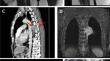
With the increasing use of next generation sequencing in soft tissue pathology, particularly in neoplasms not fitting any World Health Organization (WHO) category, the spectrum of EWSR1 fusion-associated soft tissue neoplasms has been expanding significantly. Although recurrent EWSR1::ATF1 fusions were initially limited to a triad of mesenchymal neoplasms including clear cell sarcoma of soft tissue, angiomatoid fibrous histiocytoma and malignant gastrointestinal neuroectodermal tumor (MGNET), this family has been expanding. We herein describe 4 unclassified extra-abdominal soft tissue (n = 3) and bone (n = 1) neoplasms displaying epithelioid and round cell morphology and carrying an EWSR1::ATF1 fusion. Affected were 3 males and 1 female aged 20-56 years. All primary tumors were extra-abdominal and deep-seated (chest wall, mediastinum, deltoid, and parapharyngeal soft tissue). Their size ranged 4.4-7.5 cm (median, 6.2). One patient presented with constitutional symptoms. Surgery with (2) or without (1) neo/adjuvant therapy was the treatment. At last follow-up (8-21 months), 2 patients developed progressive disease (1 recurrence; 1 distant metastasis). The immunophenotype of these tumors is potentially misleading with variable expression of EMA (2 of 3), pankeratin (2 of 4), synaptophysin (2 of 3), MUC4 (1 of 3), and ALK (1 of 3). All tumors were negative for S100 and SOX10. These observations point to the existence of heretofore under-recognized group of epithelioid and round cell neoplasms of soft tissue and bone, driven by EWSR1::ATF1 fusions, but distinct from established EWSR1::ATF1-associated soft tissue entities. Their overall morphology and immunophenotype recapitulate that of the emerging EWSR1/FUS::CREB fusion associated intra-abdominal epithelioid/round cell neoplasms. Our cases point to a potentially aggressive clinical behavior. Recognizing this tumor type is mandatory to delineate any inherent biological and/or therapeutic distinctness from other, better-known sarcomas in the differential diagnosis including sclerosing epithelioid fibrosarcoma.
期刊介绍:
Manuscripts of original studies reinforcing the evidence base of modern diagnostic pathology, using immunocytochemical, molecular and ultrastructural techniques, will be welcomed. In addition, papers on critical evaluation of diagnostic criteria but also broadsheets and guidelines with a solid evidence base will be considered. Consideration will also be given to reports of work in other fields relevant to the understanding of human pathology as well as manuscripts on the application of new methods and techniques in pathology. Submission of purely experimental articles is discouraged but manuscripts on experimental work applicable to diagnostic pathology are welcomed. Biomarker studies are welcomed but need to abide by strict rules (e.g. REMARK) of adequate sample size and relevant marker choice. Single marker studies on limited patient series without validated application will as a rule not be considered. Case reports will only be considered when they provide substantial new information with an impact on understanding disease or diagnostic practice.


 分享
分享
 求助内容:
求助内容: 应助结果提醒方式:
 扫码关注我们
