Arpitha Ravi, Philipp Bernhardt, Mathis Hoffmann, Richard Obler, Cuong Nguyen, Andreas Berting, René Chapot, Andreas Maier
{"title":"优化神经介入手术:栓塞线圈检测和自动准直以减少剂量的算法。","authors":"Arpitha Ravi, Philipp Bernhardt, Mathis Hoffmann, Richard Obler, Cuong Nguyen, Andreas Berting, René Chapot, Andreas Maier","doi":"10.1117/1.JMI.11.4.044003","DOIUrl":null,"url":null,"abstract":"<p><strong>Purpose: </strong>Monitoring radiation dose and time parameters during radiological interventions is crucial, especially in neurointerventional procedures, such as aneurysm treatment with embolization coils. The algorithm presented detects the presence of these embolization coils in medical images. It establishes a bounding box as a reference for automated collimation, with the primary objective being to enhance the efficiency and safety of neurointerventional procedures by actively optimizing image quality while minimizing patient dose.</p><p><strong>Methods: </strong>Two distinct methodologies are evaluated in our study. The first involves deep learning, employing the Faster R-CNN model with a ResNet-50 FPN as a backbone and a RetinaNet model. The second method utilizes a classical blob detection approach, serving as a benchmark for comparison.</p><p><strong>Results: </strong>We performed a fivefold cross-validation, and our top-performing model achieved mean mAP@75 of 0.84 across all folds on validation data and mean mAP@75 of 0.94 on independent test data. Since we use an upscaled bounding box, achieving 100% overlap between ground truth and prediction is not necessary. To highlight the real-world applications of our algorithm, we conducted a simulation featuring a coil constructed from an alloy wire, effectively showcasing the implementation of automatic collimation. This resulted in a notable reduction in the dose area product, signifying the reduction of stochastic risks for both patients and medical staff by minimizing scatter radiation. Additionally, our algorithm assists in avoiding extreme brightness or darkness in X-ray angiography images during narrow collimation, ultimately streamlining the collimation process for physicians.</p><p><strong>Conclusion: </strong>To our knowledge, this marks the initial attempt at an approach successfully detecting embolization coils, showcasing the extended applications of integrating detection results into the X-ray angiography system. The method we present has the potential for broader application, allowing its extension to detect other medical objects utilized in interventional procedures.</p>","PeriodicalId":47707,"journal":{"name":"Journal of Medical Imaging","volume":"11 4","pages":"044003"},"PeriodicalIF":2.3000,"publicationDate":"2024-07-01","publicationTypes":"Journal Article","fieldsOfStudy":null,"isOpenAccess":false,"openAccessPdf":"https://www.ncbi.nlm.nih.gov/pmc/articles/PMC11259374/pdf/","citationCount":"0","resultStr":"{\"title\":\"Optimizing neurointerventional procedures: an algorithm for embolization coil detection and automated collimation to enable dose reduction.\",\"authors\":\"Arpitha Ravi, Philipp Bernhardt, Mathis Hoffmann, Richard Obler, Cuong Nguyen, Andreas Berting, René Chapot, Andreas Maier\",\"doi\":\"10.1117/1.JMI.11.4.044003\",\"DOIUrl\":null,\"url\":null,\"abstract\":\"<p><strong>Purpose: </strong>Monitoring radiation dose and time parameters during radiological interventions is crucial, especially in neurointerventional procedures, such as aneurysm treatment with embolization coils. The algorithm presented detects the presence of these embolization coils in medical images. It establishes a bounding box as a reference for automated collimation, with the primary objective being to enhance the efficiency and safety of neurointerventional procedures by actively optimizing image quality while minimizing patient dose.</p><p><strong>Methods: </strong>Two distinct methodologies are evaluated in our study. The first involves deep learning, employing the Faster R-CNN model with a ResNet-50 FPN as a backbone and a RetinaNet model. The second method utilizes a classical blob detection approach, serving as a benchmark for comparison.</p><p><strong>Results: </strong>We performed a fivefold cross-validation, and our top-performing model achieved mean mAP@75 of 0.84 across all folds on validation data and mean mAP@75 of 0.94 on independent test data. Since we use an upscaled bounding box, achieving 100% overlap between ground truth and prediction is not necessary. To highlight the real-world applications of our algorithm, we conducted a simulation featuring a coil constructed from an alloy wire, effectively showcasing the implementation of automatic collimation. This resulted in a notable reduction in the dose area product, signifying the reduction of stochastic risks for both patients and medical staff by minimizing scatter radiation. Additionally, our algorithm assists in avoiding extreme brightness or darkness in X-ray angiography images during narrow collimation, ultimately streamlining the collimation process for physicians.</p><p><strong>Conclusion: </strong>To our knowledge, this marks the initial attempt at an approach successfully detecting embolization coils, showcasing the extended applications of integrating detection results into the X-ray angiography system. The method we present has the potential for broader application, allowing its extension to detect other medical objects utilized in interventional procedures.</p>\",\"PeriodicalId\":47707,\"journal\":{\"name\":\"Journal of Medical Imaging\",\"volume\":\"11 4\",\"pages\":\"044003\"},\"PeriodicalIF\":2.3000,\"publicationDate\":\"2024-07-01\",\"publicationTypes\":\"Journal Article\",\"fieldsOfStudy\":null,\"isOpenAccess\":false,\"openAccessPdf\":\"https://www.ncbi.nlm.nih.gov/pmc/articles/PMC11259374/pdf/\",\"citationCount\":\"0\",\"resultStr\":null,\"platform\":\"Semanticscholar\",\"paperid\":null,\"PeriodicalName\":\"Journal of Medical Imaging\",\"FirstCategoryId\":\"3\",\"ListUrlMain\":\"https://doi.org/10.1117/1.JMI.11.4.044003\",\"RegionNum\":0,\"RegionCategory\":null,\"ArticlePicture\":[],\"TitleCN\":null,\"AbstractTextCN\":null,\"PMCID\":null,\"EPubDate\":\"2024/7/17 0:00:00\",\"PubModel\":\"Epub\",\"JCR\":\"Q3\",\"JCRName\":\"RADIOLOGY, NUCLEAR MEDICINE & MEDICAL IMAGING\",\"Score\":null,\"Total\":0}","platform":"Semanticscholar","paperid":null,"PeriodicalName":"Journal of Medical Imaging","FirstCategoryId":"3","ListUrlMain":"https://doi.org/10.1117/1.JMI.11.4.044003","RegionNum":0,"RegionCategory":null,"ArticlePicture":[],"TitleCN":null,"AbstractTextCN":null,"PMCID":null,"EPubDate":"2024/7/17 0:00:00","PubModel":"Epub","JCR":"Q3","JCRName":"RADIOLOGY, NUCLEAR MEDICINE & MEDICAL IMAGING","Score":null,"Total":0}
Optimizing neurointerventional procedures: an algorithm for embolization coil detection and automated collimation to enable dose reduction.
Purpose: Monitoring radiation dose and time parameters during radiological interventions is crucial, especially in neurointerventional procedures, such as aneurysm treatment with embolization coils. The algorithm presented detects the presence of these embolization coils in medical images. It establishes a bounding box as a reference for automated collimation, with the primary objective being to enhance the efficiency and safety of neurointerventional procedures by actively optimizing image quality while minimizing patient dose.
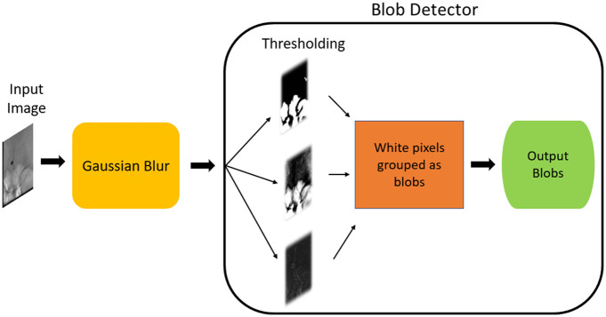
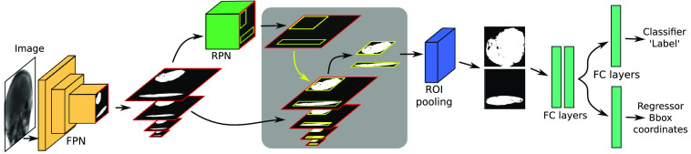
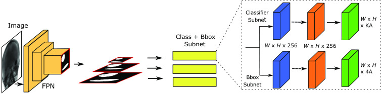
Methods: Two distinct methodologies are evaluated in our study. The first involves deep learning, employing the Faster R-CNN model with a ResNet-50 FPN as a backbone and a RetinaNet model. The second method utilizes a classical blob detection approach, serving as a benchmark for comparison.
Results: We performed a fivefold cross-validation, and our top-performing model achieved mean mAP@75 of 0.84 across all folds on validation data and mean mAP@75 of 0.94 on independent test data. Since we use an upscaled bounding box, achieving 100% overlap between ground truth and prediction is not necessary. To highlight the real-world applications of our algorithm, we conducted a simulation featuring a coil constructed from an alloy wire, effectively showcasing the implementation of automatic collimation. This resulted in a notable reduction in the dose area product, signifying the reduction of stochastic risks for both patients and medical staff by minimizing scatter radiation. Additionally, our algorithm assists in avoiding extreme brightness or darkness in X-ray angiography images during narrow collimation, ultimately streamlining the collimation process for physicians.
Conclusion: To our knowledge, this marks the initial attempt at an approach successfully detecting embolization coils, showcasing the extended applications of integrating detection results into the X-ray angiography system. The method we present has the potential for broader application, allowing its extension to detect other medical objects utilized in interventional procedures.
期刊介绍:
JMI covers fundamental and translational research, as well as applications, focused on medical imaging, which continue to yield physical and biomedical advancements in the early detection, diagnostics, and therapy of disease as well as in the understanding of normal. The scope of JMI includes: Imaging physics, Tomographic reconstruction algorithms (such as those in CT and MRI), Image processing and deep learning, Computer-aided diagnosis and quantitative image analysis, Visualization and modeling, Picture archiving and communications systems (PACS), Image perception and observer performance, Technology assessment, Ultrasonic imaging, Image-guided procedures, Digital pathology, Biomedical applications of biomedical imaging. JMI allows for the peer-reviewed communication and archiving of scientific developments, translational and clinical applications, reviews, and recommendations for the field.


 分享
分享
 求助内容:
求助内容: 应助结果提醒方式:
 扫码关注我们
