{"title":"将眼球运动功能评估作为轻度脑外伤的生物标志物。","authors":"Ekaterina Lunkova, Jen-Kai Chen, Rajeet Singh Saluja, Alain Ptito","doi":"10.1089/neur.2024.0018","DOIUrl":null,"url":null,"abstract":"<p><p>Mild traumatic brain injury (mTBI), or concussion, is a major public health problem, and ambiguity still exists regarding its diagnosis. While functional magnetic resonance imaging (fMRI) has been identified as a helpful screening tool for concussion, its limited accessibility in clinical or field settings necessitates a more efficient alternative. Oculomotor function deficit is an often-reported pathology in mTBI. Due to the neuroanatomical overlap between eye-movement circuitry and mTBI pathophysiology, visual deficits are expected. In this study, we investigate the possibility of using an oculomotor assessment tool for finding biomarkers in concussion. We used fMRI with tasks evaluating oculomotor functions: smooth pursuit (SP), saccades, anti-saccades, and optokinetic nystagmus (OKN). Before the scanning, the testing with a system of virtual reality goggles with integrated eye- and head-tracking was used where subjects performed the same tasks as those used in fMRI. Twenty-nine concussed symptomatic adults (CSA) within 1-month postconcussion and 29 age- and sex-matched healthy controls (HCS) were tested to examine blood oxygen level-dependent (BOLD) fMRI alterations associated with performances in oculomotor function after mTBI and evaluate the efficacy of the oculomotor assessment in detecting oculomotor and gaze deficits following mTBI. Comparing CSA with HCS, significant differences were observed in anti-saccades and OKN performance. CSA group exhibited elevated %BOLD signal change on each task compared with HCS: in the superior frontal gyrus during the smooth pursuit, inferior frontal gyrus during the saccades, putamen and dorsolateral prefrontal cortex (DLPFC) during the anti-saccades, and lingual gyrus and IFG during the OKN. Key findings include the following: (1) oculomotor deficits in concussed subjects compared with controls, (2) abnormal activation patterns in areas related to the regulation and control of oculomotor movements, suggesting concussion-induced disruptions, and (3) the potential of oculomotor assessment as a promising approach for mTBI biomarkers, with anti-saccades and OKN identified as the most sensitive tasks.</p>","PeriodicalId":74300,"journal":{"name":"Neurotrauma reports","volume":"5 1","pages":"628-639"},"PeriodicalIF":1.8000,"publicationDate":"2024-07-03","publicationTypes":"Journal Article","fieldsOfStudy":null,"isOpenAccess":false,"openAccessPdf":"https://www.ncbi.nlm.nih.gov/pmc/articles/PMC11257115/pdf/","citationCount":"0","resultStr":"{\"title\":\"Assessment of Oculomotor Functions as a Biomarker in Mild Traumatic Brain Injury.\",\"authors\":\"Ekaterina Lunkova, Jen-Kai Chen, Rajeet Singh Saluja, Alain Ptito\",\"doi\":\"10.1089/neur.2024.0018\",\"DOIUrl\":null,\"url\":null,\"abstract\":\"<p><p>Mild traumatic brain injury (mTBI), or concussion, is a major public health problem, and ambiguity still exists regarding its diagnosis. While functional magnetic resonance imaging (fMRI) has been identified as a helpful screening tool for concussion, its limited accessibility in clinical or field settings necessitates a more efficient alternative. Oculomotor function deficit is an often-reported pathology in mTBI. Due to the neuroanatomical overlap between eye-movement circuitry and mTBI pathophysiology, visual deficits are expected. In this study, we investigate the possibility of using an oculomotor assessment tool for finding biomarkers in concussion. We used fMRI with tasks evaluating oculomotor functions: smooth pursuit (SP), saccades, anti-saccades, and optokinetic nystagmus (OKN). Before the scanning, the testing with a system of virtual reality goggles with integrated eye- and head-tracking was used where subjects performed the same tasks as those used in fMRI. Twenty-nine concussed symptomatic adults (CSA) within 1-month postconcussion and 29 age- and sex-matched healthy controls (HCS) were tested to examine blood oxygen level-dependent (BOLD) fMRI alterations associated with performances in oculomotor function after mTBI and evaluate the efficacy of the oculomotor assessment in detecting oculomotor and gaze deficits following mTBI. Comparing CSA with HCS, significant differences were observed in anti-saccades and OKN performance. CSA group exhibited elevated %BOLD signal change on each task compared with HCS: in the superior frontal gyrus during the smooth pursuit, inferior frontal gyrus during the saccades, putamen and dorsolateral prefrontal cortex (DLPFC) during the anti-saccades, and lingual gyrus and IFG during the OKN. Key findings include the following: (1) oculomotor deficits in concussed subjects compared with controls, (2) abnormal activation patterns in areas related to the regulation and control of oculomotor movements, suggesting concussion-induced disruptions, and (3) the potential of oculomotor assessment as a promising approach for mTBI biomarkers, with anti-saccades and OKN identified as the most sensitive tasks.</p>\",\"PeriodicalId\":74300,\"journal\":{\"name\":\"Neurotrauma reports\",\"volume\":\"5 1\",\"pages\":\"628-639\"},\"PeriodicalIF\":1.8000,\"publicationDate\":\"2024-07-03\",\"publicationTypes\":\"Journal Article\",\"fieldsOfStudy\":null,\"isOpenAccess\":false,\"openAccessPdf\":\"https://www.ncbi.nlm.nih.gov/pmc/articles/PMC11257115/pdf/\",\"citationCount\":\"0\",\"resultStr\":null,\"platform\":\"Semanticscholar\",\"paperid\":null,\"PeriodicalName\":\"Neurotrauma reports\",\"FirstCategoryId\":\"1085\",\"ListUrlMain\":\"https://doi.org/10.1089/neur.2024.0018\",\"RegionNum\":0,\"RegionCategory\":null,\"ArticlePicture\":[],\"TitleCN\":null,\"AbstractTextCN\":null,\"PMCID\":null,\"EPubDate\":\"2024/1/1 0:00:00\",\"PubModel\":\"eCollection\",\"JCR\":\"Q3\",\"JCRName\":\"CLINICAL NEUROLOGY\",\"Score\":null,\"Total\":0}","platform":"Semanticscholar","paperid":null,"PeriodicalName":"Neurotrauma reports","FirstCategoryId":"1085","ListUrlMain":"https://doi.org/10.1089/neur.2024.0018","RegionNum":0,"RegionCategory":null,"ArticlePicture":[],"TitleCN":null,"AbstractTextCN":null,"PMCID":null,"EPubDate":"2024/1/1 0:00:00","PubModel":"eCollection","JCR":"Q3","JCRName":"CLINICAL NEUROLOGY","Score":null,"Total":0}
Assessment of Oculomotor Functions as a Biomarker in Mild Traumatic Brain Injury.
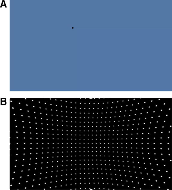
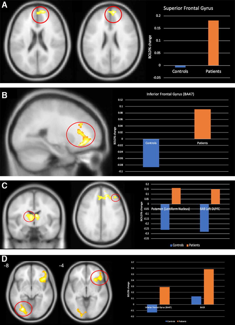
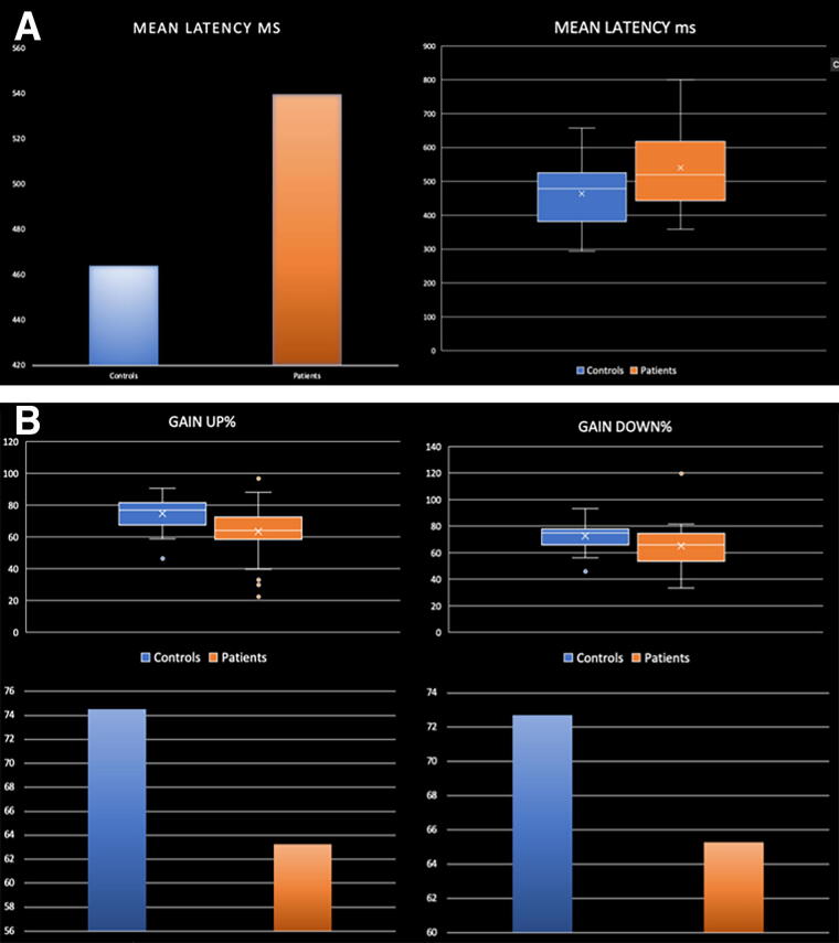
Mild traumatic brain injury (mTBI), or concussion, is a major public health problem, and ambiguity still exists regarding its diagnosis. While functional magnetic resonance imaging (fMRI) has been identified as a helpful screening tool for concussion, its limited accessibility in clinical or field settings necessitates a more efficient alternative. Oculomotor function deficit is an often-reported pathology in mTBI. Due to the neuroanatomical overlap between eye-movement circuitry and mTBI pathophysiology, visual deficits are expected. In this study, we investigate the possibility of using an oculomotor assessment tool for finding biomarkers in concussion. We used fMRI with tasks evaluating oculomotor functions: smooth pursuit (SP), saccades, anti-saccades, and optokinetic nystagmus (OKN). Before the scanning, the testing with a system of virtual reality goggles with integrated eye- and head-tracking was used where subjects performed the same tasks as those used in fMRI. Twenty-nine concussed symptomatic adults (CSA) within 1-month postconcussion and 29 age- and sex-matched healthy controls (HCS) were tested to examine blood oxygen level-dependent (BOLD) fMRI alterations associated with performances in oculomotor function after mTBI and evaluate the efficacy of the oculomotor assessment in detecting oculomotor and gaze deficits following mTBI. Comparing CSA with HCS, significant differences were observed in anti-saccades and OKN performance. CSA group exhibited elevated %BOLD signal change on each task compared with HCS: in the superior frontal gyrus during the smooth pursuit, inferior frontal gyrus during the saccades, putamen and dorsolateral prefrontal cortex (DLPFC) during the anti-saccades, and lingual gyrus and IFG during the OKN. Key findings include the following: (1) oculomotor deficits in concussed subjects compared with controls, (2) abnormal activation patterns in areas related to the regulation and control of oculomotor movements, suggesting concussion-induced disruptions, and (3) the potential of oculomotor assessment as a promising approach for mTBI biomarkers, with anti-saccades and OKN identified as the most sensitive tasks.


 分享
分享

 求助内容:
求助内容: 应助结果提醒方式:
 扫码关注我们
