{"title":"超声引导下沿肱骨定位桡神经:为更安全的上臂手术提供参考点。","authors":"T Da Silva, D Mueck, C Knop, T Merkle","doi":"10.1007/s12306-024-00841-1","DOIUrl":null,"url":null,"abstract":"<p><strong>Purpose: </strong>The close proximity of the radial nerve to the humerus poses a risk during upper arm surgery. Although the general course of the radial nerve is well-known, its exact position in relation to anatomical reference points remains poorly investigated. This study aimed to develop a standardized protocol for the sonographic and clinical identification of the radial nerve in the upper arm. The ultimate goal is to assist surgeons in avoiding iatrogenic radial nerve palsy.</p><p><strong>Methods: </strong>A total of 76 measurements were performed in 38 volunteers (both sides). Ultrasound measurements were performed using a linear transducer (10 MHz) to identify the radial nerve at two key points: RD (where the radial nerve crosses the dorsal surface of the humerus) and RL (where the radial nerve crosses the lateral aspect of the humerus). Distances from specific reference points (acromion, lateral epicondyle, medial epicondyle, olecranon fossa) to RD and RL were measured, and the angle between the course of the nerve and the humeral axis was recorded. Humeral length was defined as the distance between the posterodorsal corner of the acromion and the lateral epicondyle.</p><p><strong>Results: </strong>The distance from the lateral epicondyle to RD was on average 15.5 cm ± 1.3, corresponding to 50% of the humeral length. The distance from the lateral epicondyle to RL was on average 6.7 cm ± 0.8, corresponding to 21% of the humeral length. The course of the nerve between RD and RL showed an average angulation of 37° to the anatomical axis of the humerus. Gender, BMI, dominant hand, and arm thickness did not correlate with the distances to RD or RL. Measurements were consistent between the left and right side.</p><p><strong>Conclusion: </strong>The radial nerve can typically be identified by employing a 1/2 and 1/5 ratio on the dorsal and lateral aspects of the humerus. Due to slight variations in individual anatomy, the utilization of ultrasound-assisted visualization presents a valuable and straightforward approach to mitigate the risk of iatrogenic radial nerve palsy during upper arm surgery. This study introduces an easy and fast protocol for this purpose.</p>","PeriodicalId":18875,"journal":{"name":"MUSCULOSKELETAL SURGERY","volume":" ","pages":"47-53"},"PeriodicalIF":0.0000,"publicationDate":"2025-03-01","publicationTypes":"Journal Article","fieldsOfStudy":null,"isOpenAccess":false,"openAccessPdf":"https://www.ncbi.nlm.nih.gov/pmc/articles/PMC11876194/pdf/","citationCount":"0","resultStr":"{\"title\":\"Ultrasound-guided localization of the radial nerve along the humerus: providing reference points for safer upper arm surgery.\",\"authors\":\"T Da Silva, D Mueck, C Knop, T Merkle\",\"doi\":\"10.1007/s12306-024-00841-1\",\"DOIUrl\":null,\"url\":null,\"abstract\":\"<p><strong>Purpose: </strong>The close proximity of the radial nerve to the humerus poses a risk during upper arm surgery. Although the general course of the radial nerve is well-known, its exact position in relation to anatomical reference points remains poorly investigated. This study aimed to develop a standardized protocol for the sonographic and clinical identification of the radial nerve in the upper arm. The ultimate goal is to assist surgeons in avoiding iatrogenic radial nerve palsy.</p><p><strong>Methods: </strong>A total of 76 measurements were performed in 38 volunteers (both sides). Ultrasound measurements were performed using a linear transducer (10 MHz) to identify the radial nerve at two key points: RD (where the radial nerve crosses the dorsal surface of the humerus) and RL (where the radial nerve crosses the lateral aspect of the humerus). Distances from specific reference points (acromion, lateral epicondyle, medial epicondyle, olecranon fossa) to RD and RL were measured, and the angle between the course of the nerve and the humeral axis was recorded. Humeral length was defined as the distance between the posterodorsal corner of the acromion and the lateral epicondyle.</p><p><strong>Results: </strong>The distance from the lateral epicondyle to RD was on average 15.5 cm ± 1.3, corresponding to 50% of the humeral length. The distance from the lateral epicondyle to RL was on average 6.7 cm ± 0.8, corresponding to 21% of the humeral length. The course of the nerve between RD and RL showed an average angulation of 37° to the anatomical axis of the humerus. Gender, BMI, dominant hand, and arm thickness did not correlate with the distances to RD or RL. Measurements were consistent between the left and right side.</p><p><strong>Conclusion: </strong>The radial nerve can typically be identified by employing a 1/2 and 1/5 ratio on the dorsal and lateral aspects of the humerus. Due to slight variations in individual anatomy, the utilization of ultrasound-assisted visualization presents a valuable and straightforward approach to mitigate the risk of iatrogenic radial nerve palsy during upper arm surgery. This study introduces an easy and fast protocol for this purpose.</p>\",\"PeriodicalId\":18875,\"journal\":{\"name\":\"MUSCULOSKELETAL SURGERY\",\"volume\":\" \",\"pages\":\"47-53\"},\"PeriodicalIF\":0.0000,\"publicationDate\":\"2025-03-01\",\"publicationTypes\":\"Journal Article\",\"fieldsOfStudy\":null,\"isOpenAccess\":false,\"openAccessPdf\":\"https://www.ncbi.nlm.nih.gov/pmc/articles/PMC11876194/pdf/\",\"citationCount\":\"0\",\"resultStr\":null,\"platform\":\"Semanticscholar\",\"paperid\":null,\"PeriodicalName\":\"MUSCULOSKELETAL SURGERY\",\"FirstCategoryId\":\"1085\",\"ListUrlMain\":\"https://doi.org/10.1007/s12306-024-00841-1\",\"RegionNum\":0,\"RegionCategory\":null,\"ArticlePicture\":[],\"TitleCN\":null,\"AbstractTextCN\":null,\"PMCID\":null,\"EPubDate\":\"2024/7/23 0:00:00\",\"PubModel\":\"Epub\",\"JCR\":\"Q1\",\"JCRName\":\"Medicine\",\"Score\":null,\"Total\":0}","platform":"Semanticscholar","paperid":null,"PeriodicalName":"MUSCULOSKELETAL SURGERY","FirstCategoryId":"1085","ListUrlMain":"https://doi.org/10.1007/s12306-024-00841-1","RegionNum":0,"RegionCategory":null,"ArticlePicture":[],"TitleCN":null,"AbstractTextCN":null,"PMCID":null,"EPubDate":"2024/7/23 0:00:00","PubModel":"Epub","JCR":"Q1","JCRName":"Medicine","Score":null,"Total":0}
Ultrasound-guided localization of the radial nerve along the humerus: providing reference points for safer upper arm surgery.
Purpose: The close proximity of the radial nerve to the humerus poses a risk during upper arm surgery. Although the general course of the radial nerve is well-known, its exact position in relation to anatomical reference points remains poorly investigated. This study aimed to develop a standardized protocol for the sonographic and clinical identification of the radial nerve in the upper arm. The ultimate goal is to assist surgeons in avoiding iatrogenic radial nerve palsy.
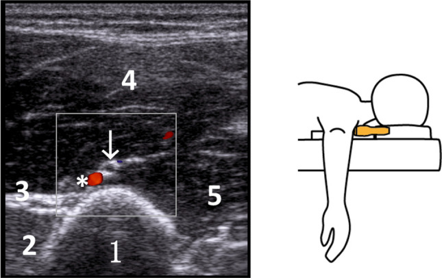
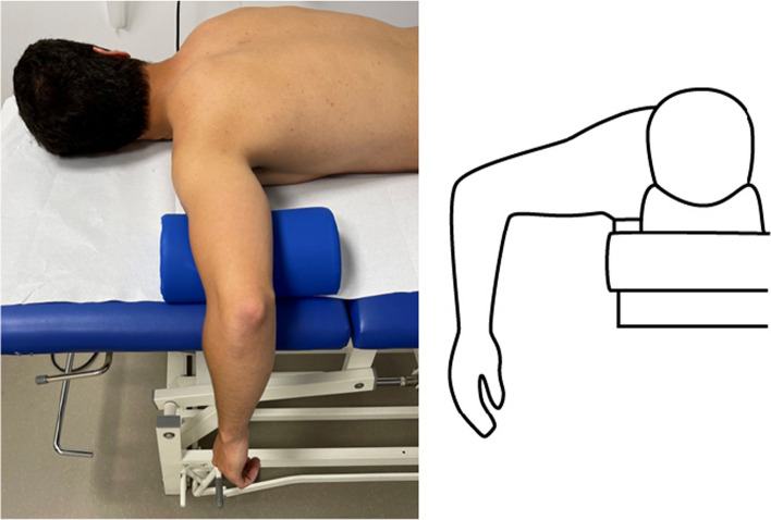
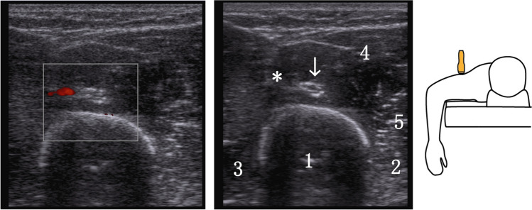
Methods: A total of 76 measurements were performed in 38 volunteers (both sides). Ultrasound measurements were performed using a linear transducer (10 MHz) to identify the radial nerve at two key points: RD (where the radial nerve crosses the dorsal surface of the humerus) and RL (where the radial nerve crosses the lateral aspect of the humerus). Distances from specific reference points (acromion, lateral epicondyle, medial epicondyle, olecranon fossa) to RD and RL were measured, and the angle between the course of the nerve and the humeral axis was recorded. Humeral length was defined as the distance between the posterodorsal corner of the acromion and the lateral epicondyle.
Results: The distance from the lateral epicondyle to RD was on average 15.5 cm ± 1.3, corresponding to 50% of the humeral length. The distance from the lateral epicondyle to RL was on average 6.7 cm ± 0.8, corresponding to 21% of the humeral length. The course of the nerve between RD and RL showed an average angulation of 37° to the anatomical axis of the humerus. Gender, BMI, dominant hand, and arm thickness did not correlate with the distances to RD or RL. Measurements were consistent between the left and right side.
Conclusion: The radial nerve can typically be identified by employing a 1/2 and 1/5 ratio on the dorsal and lateral aspects of the humerus. Due to slight variations in individual anatomy, the utilization of ultrasound-assisted visualization presents a valuable and straightforward approach to mitigate the risk of iatrogenic radial nerve palsy during upper arm surgery. This study introduces an easy and fast protocol for this purpose.
期刊介绍:
Musculoskeletal Surgery – Formerly La Chirurgia degli Organi di Movimento, founded in 1917 at the Istituto Ortopedico Rizzoli, is a peer-reviewed journal published three times a year. The journal provides up-to-date information to clinicians and scientists through the publication of original papers, reviews, case reports, and brief communications dealing with the pathogenesis and treatment of orthopaedic conditions.An electronic version is also available at http://www.springerlink.com.The journal is open for publication of supplements and for publishing abstracts of scientific meetings; conditions can be obtained from the Editors-in-Chief or the Publisher.


 分享
分享
 求助内容:
求助内容: 应助结果提醒方式:
 扫码关注我们
