Amy R. McDowell, Laura Zambreanu, Hamza A. Salhab, Carolynne M. Doherty, Philippa Bridgen, Pete Lally, Sachit Shah, Zimu Huo, Stephen J. Wastling, Tarek Yousry, Jasper Morrow, John S. Thornton, Michael P. Lunn
{"title":"使用高分辨率磁共振成像在 7 特斯拉下观察健康志愿者的硬脊膜神经和周围解剖结构的初步发现。","authors":"Amy R. McDowell, Laura Zambreanu, Hamza A. Salhab, Carolynne M. Doherty, Philippa Bridgen, Pete Lally, Sachit Shah, Zimu Huo, Stephen J. Wastling, Tarek Yousry, Jasper Morrow, John S. Thornton, Michael P. Lunn","doi":"10.1111/jns.12645","DOIUrl":null,"url":null,"abstract":"<div>\n \n \n <section>\n \n <h3> Background and Aims</h3>\n \n <p>Histopathological diagnosis is the gold standard in many acquired inflammatory, infiltrative and amyloid based peripheral nerve diseases and a sensory nerve biopsy of sural or superficial peroneal nerve is favoured where a biopsy is deemed necessary. The ability to determine nerve pathology by high-resolution imaging techniques resolving anatomy and imaging characteristics might improve diagnosis and obviate the need for biopsy in some. The sural nerve is anatomically variable and occasionally adjacent vessels can be sent for analysis in error. Knowing the exact position and relationships of the nerve prior to surgery could be clinically useful and thus reliably resolving nerve position has some utility.</p>\n </section>\n \n <section>\n \n <h3> Methods</h3>\n \n <p>7T images of eight healthy volunteers' (HV) right ankle were acquired in a pilot study using a double-echo in steady-state sequence for high-resolution anatomy images. Magnetic Transfer Ratio images were acquired of the same area. Systematic scoring of the sural, tibial and deep peroneal nerve around the surgical landmark 7 cm from the lateral malleolus was performed (number of fascicles, area in voxels and mm<sup>2</sup>, diameter and location relative to nearby vessels and muscles).</p>\n </section>\n \n <section>\n \n <h3> Results</h3>\n \n <p>The sural and tibial nerves were visualised in the high-resolution double-echo in steady-state (DESS) image in all HV. The deep peroneal nerve was not always visualised at level of interest. The MTR values were tightly grouped except in the sural nerve where the nerve was not visualised in two HV. The sural nerve location was found to be variable (e.g., lateral or medial to, or crossing behind, or found positioned directly posterior to the saphenous vein).</p>\n </section>\n \n <section>\n \n <h3> Interpretation</h3>\n \n <p>High-resolution high-field images have excellent visualisation of the sural nerve and would give surgeons prior knowledge of the position before surgery. Basic imaging characteristics of the sural nerve can be acquired, but more detailed imaging characteristics are not easily evaluable in the very small sural and further developments and specific studies are required for any diagnostic utility at 7T.</p>\n </section>\n </div>","PeriodicalId":17451,"journal":{"name":"Journal of the Peripheral Nervous System","volume":"29 3","pages":"368-375"},"PeriodicalIF":3.0000,"publicationDate":"2024-07-26","publicationTypes":"Journal Article","fieldsOfStudy":null,"isOpenAccess":false,"openAccessPdf":"https://onlinelibrary.wiley.com/doi/epdf/10.1111/jns.12645","citationCount":"0","resultStr":"{\"title\":\"Initial findings using high-resolution magnetic resonance imaging for visualisation of the sural nerve and surrounding anatomy in healthy volunteers at 7 Tesla\",\"authors\":\"Amy R. McDowell, Laura Zambreanu, Hamza A. Salhab, Carolynne M. Doherty, Philippa Bridgen, Pete Lally, Sachit Shah, Zimu Huo, Stephen J. Wastling, Tarek Yousry, Jasper Morrow, John S. Thornton, Michael P. Lunn\",\"doi\":\"10.1111/jns.12645\",\"DOIUrl\":null,\"url\":null,\"abstract\":\"<div>\\n \\n \\n <section>\\n \\n <h3> Background and Aims</h3>\\n \\n <p>Histopathological diagnosis is the gold standard in many acquired inflammatory, infiltrative and amyloid based peripheral nerve diseases and a sensory nerve biopsy of sural or superficial peroneal nerve is favoured where a biopsy is deemed necessary. The ability to determine nerve pathology by high-resolution imaging techniques resolving anatomy and imaging characteristics might improve diagnosis and obviate the need for biopsy in some. The sural nerve is anatomically variable and occasionally adjacent vessels can be sent for analysis in error. Knowing the exact position and relationships of the nerve prior to surgery could be clinically useful and thus reliably resolving nerve position has some utility.</p>\\n </section>\\n \\n <section>\\n \\n <h3> Methods</h3>\\n \\n <p>7T images of eight healthy volunteers' (HV) right ankle were acquired in a pilot study using a double-echo in steady-state sequence for high-resolution anatomy images. Magnetic Transfer Ratio images were acquired of the same area. Systematic scoring of the sural, tibial and deep peroneal nerve around the surgical landmark 7 cm from the lateral malleolus was performed (number of fascicles, area in voxels and mm<sup>2</sup>, diameter and location relative to nearby vessels and muscles).</p>\\n </section>\\n \\n <section>\\n \\n <h3> Results</h3>\\n \\n <p>The sural and tibial nerves were visualised in the high-resolution double-echo in steady-state (DESS) image in all HV. The deep peroneal nerve was not always visualised at level of interest. The MTR values were tightly grouped except in the sural nerve where the nerve was not visualised in two HV. The sural nerve location was found to be variable (e.g., lateral or medial to, or crossing behind, or found positioned directly posterior to the saphenous vein).</p>\\n </section>\\n \\n <section>\\n \\n <h3> Interpretation</h3>\\n \\n <p>High-resolution high-field images have excellent visualisation of the sural nerve and would give surgeons prior knowledge of the position before surgery. Basic imaging characteristics of the sural nerve can be acquired, but more detailed imaging characteristics are not easily evaluable in the very small sural and further developments and specific studies are required for any diagnostic utility at 7T.</p>\\n </section>\\n </div>\",\"PeriodicalId\":17451,\"journal\":{\"name\":\"Journal of the Peripheral Nervous System\",\"volume\":\"29 3\",\"pages\":\"368-375\"},\"PeriodicalIF\":3.0000,\"publicationDate\":\"2024-07-26\",\"publicationTypes\":\"Journal Article\",\"fieldsOfStudy\":null,\"isOpenAccess\":false,\"openAccessPdf\":\"https://onlinelibrary.wiley.com/doi/epdf/10.1111/jns.12645\",\"citationCount\":\"0\",\"resultStr\":null,\"platform\":\"Semanticscholar\",\"paperid\":null,\"PeriodicalName\":\"Journal of the Peripheral Nervous System\",\"FirstCategoryId\":\"3\",\"ListUrlMain\":\"https://onlinelibrary.wiley.com/doi/10.1111/jns.12645\",\"RegionNum\":3,\"RegionCategory\":\"医学\",\"ArticlePicture\":[],\"TitleCN\":null,\"AbstractTextCN\":null,\"PMCID\":null,\"EPubDate\":\"\",\"PubModel\":\"\",\"JCR\":\"Q1\",\"JCRName\":\"CLINICAL NEUROLOGY\",\"Score\":null,\"Total\":0}","platform":"Semanticscholar","paperid":null,"PeriodicalName":"Journal of the Peripheral Nervous System","FirstCategoryId":"3","ListUrlMain":"https://onlinelibrary.wiley.com/doi/10.1111/jns.12645","RegionNum":3,"RegionCategory":"医学","ArticlePicture":[],"TitleCN":null,"AbstractTextCN":null,"PMCID":null,"EPubDate":"","PubModel":"","JCR":"Q1","JCRName":"CLINICAL NEUROLOGY","Score":null,"Total":0}
Initial findings using high-resolution magnetic resonance imaging for visualisation of the sural nerve and surrounding anatomy in healthy volunteers at 7 Tesla
Background and Aims
Histopathological diagnosis is the gold standard in many acquired inflammatory, infiltrative and amyloid based peripheral nerve diseases and a sensory nerve biopsy of sural or superficial peroneal nerve is favoured where a biopsy is deemed necessary. The ability to determine nerve pathology by high-resolution imaging techniques resolving anatomy and imaging characteristics might improve diagnosis and obviate the need for biopsy in some. The sural nerve is anatomically variable and occasionally adjacent vessels can be sent for analysis in error. Knowing the exact position and relationships of the nerve prior to surgery could be clinically useful and thus reliably resolving nerve position has some utility.
Methods
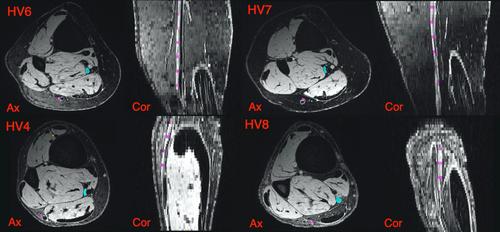
7T images of eight healthy volunteers' (HV) right ankle were acquired in a pilot study using a double-echo in steady-state sequence for high-resolution anatomy images. Magnetic Transfer Ratio images were acquired of the same area. Systematic scoring of the sural, tibial and deep peroneal nerve around the surgical landmark 7 cm from the lateral malleolus was performed (number of fascicles, area in voxels and mm2, diameter and location relative to nearby vessels and muscles).
Results
The sural and tibial nerves were visualised in the high-resolution double-echo in steady-state (DESS) image in all HV. The deep peroneal nerve was not always visualised at level of interest. The MTR values were tightly grouped except in the sural nerve where the nerve was not visualised in two HV. The sural nerve location was found to be variable (e.g., lateral or medial to, or crossing behind, or found positioned directly posterior to the saphenous vein).
Interpretation
High-resolution high-field images have excellent visualisation of the sural nerve and would give surgeons prior knowledge of the position before surgery. Basic imaging characteristics of the sural nerve can be acquired, but more detailed imaging characteristics are not easily evaluable in the very small sural and further developments and specific studies are required for any diagnostic utility at 7T.
期刊介绍:
The Journal of the Peripheral Nervous System is the official journal of the Peripheral Nerve Society. Founded in 1996, it is the scientific journal of choice for clinicians, clinical scientists and basic neuroscientists interested in all aspects of biology and clinical research of peripheral nervous system disorders.
The Journal of the Peripheral Nervous System is a peer-reviewed journal that publishes high quality articles on cell and molecular biology, genomics, neuropathic pain, clinical research, trials, and unique case reports on inherited and acquired peripheral neuropathies.
Original articles are organized according to the topic in one of four specific areas: Mechanisms of Disease, Genetics, Clinical Research, and Clinical Trials.
The journal also publishes regular review papers on hot topics and Special Issues on basic, clinical, or assembled research in the field of peripheral nervous system disorders. Authors interested in contributing a review-type article or a Special Issue should contact the Editorial Office to discuss the scope of the proposed article with the Editor-in-Chief.


 分享
分享

 求助内容:
求助内容: 应助结果提醒方式:
 扫码关注我们
