Anna Bjerkén, Hanna Tomic, Sophia Zackrisson, Magnus Dustler, Predrag R Bakic, Anders Tingberg
{"title":"估算同步数字乳腺断层成像和机械成像的吸收剂量。","authors":"Anna Bjerkén, Hanna Tomic, Sophia Zackrisson, Magnus Dustler, Predrag R Bakic, Anders Tingberg","doi":"10.1117/1.JMI.12.S1.S13003","DOIUrl":null,"url":null,"abstract":"<p><strong>Purpose: </strong>Use of mechanical imaging (MI) as complementary to digital mammography (DM), or in simultaneous digital breast tomosynthesis (DBT) and MI - DBTMI, has demonstrated the potential to increase the specificity of breast cancer screening and reduce unnecessary biopsies compared with DM. The aim of this study is to investigate the increase in the radiation dose due to the presence of an MI sensor during simultaneous image acquisition when automatic exposure control is used.</p><p><strong>Approach: </strong>A radiation dose study was conducted on clinically available breast imaging systems with and without an MI sensor present. Our estimations were based on three approaches. In the first approach, exposure values were compared in paired clinical DBT and DBTMI acquisitions in 97 women. In the second approach polymethyl methacrylate (PMMA) phantoms of various thicknesses were used, and the average glandular dose (AGD) values were compared. Finally, a rectangular PMMA phantom with a 45 mm thickness was used, and the AGD values were estimated based on air kerma measurements with an electronic dosemeter.</p><p><strong>Results: </strong>The relative increase in exposure estimated from digital imaging and communications in medicine headers when using an MI sensor in clinical DBTMI was <math><mrow><mn>11.9</mn> <mo>%</mo> <mo>±</mo> <mn>10.4</mn></mrow> </math> . For the phantom measurements of various thicknesses of PMMA, the relative increases in the AGD for DM and DBT measurements were, on average, <math><mrow><mn>10.7</mn> <mo>%</mo> <mo>±</mo> <mn>3.1</mn></mrow> </math> and <math><mrow><mn>11.4</mn> <mo>%</mo> <mo>±</mo> <mn>3.0</mn></mrow> </math> , respectively. The relative increase in the AGD using the electronic dosemeter was <math><mrow><mn>11.2</mn> <mo>%</mo> <mo>±</mo> <mo><</mo> <mn>0.001</mn></mrow> </math> in DM and <math><mrow><mn>12.2</mn> <mo>%</mo> <mo>±</mo> <mo><</mo> <mn>0.001</mn></mrow> </math> in DBT. The average difference in dose between the methods was <math><mrow><mn>11.5</mn> <mo>%</mo> <mo>±</mo> <mn>3.3</mn></mrow> </math> .</p><p><strong>Conclusions: </strong>Our measurements suggest that the use of simultaneous breast radiography and MI increases the AGD by an average of <math><mrow><mn>11.5</mn> <mo>%</mo> <mo>±</mo> <mn>3.3</mn></mrow> </math> . The increase in dose is within the acceptable values for mammography screening recommended by European guidelines.</p>","PeriodicalId":47707,"journal":{"name":"Journal of Medical Imaging","volume":"12 Suppl 1","pages":"S13003"},"PeriodicalIF":2.3000,"publicationDate":"2025-01-01","publicationTypes":"Journal Article","fieldsOfStudy":null,"isOpenAccess":false,"openAccessPdf":"https://www.ncbi.nlm.nih.gov/pmc/articles/PMC11266811/pdf/","citationCount":"0","resultStr":"{\"title\":\"Estimation of the absorbed dose in simultaneous digital breast tomosynthesis and mechanical imaging.\",\"authors\":\"Anna Bjerkén, Hanna Tomic, Sophia Zackrisson, Magnus Dustler, Predrag R Bakic, Anders Tingberg\",\"doi\":\"10.1117/1.JMI.12.S1.S13003\",\"DOIUrl\":null,\"url\":null,\"abstract\":\"<p><strong>Purpose: </strong>Use of mechanical imaging (MI) as complementary to digital mammography (DM), or in simultaneous digital breast tomosynthesis (DBT) and MI - DBTMI, has demonstrated the potential to increase the specificity of breast cancer screening and reduce unnecessary biopsies compared with DM. The aim of this study is to investigate the increase in the radiation dose due to the presence of an MI sensor during simultaneous image acquisition when automatic exposure control is used.</p><p><strong>Approach: </strong>A radiation dose study was conducted on clinically available breast imaging systems with and without an MI sensor present. Our estimations were based on three approaches. In the first approach, exposure values were compared in paired clinical DBT and DBTMI acquisitions in 97 women. In the second approach polymethyl methacrylate (PMMA) phantoms of various thicknesses were used, and the average glandular dose (AGD) values were compared. Finally, a rectangular PMMA phantom with a 45 mm thickness was used, and the AGD values were estimated based on air kerma measurements with an electronic dosemeter.</p><p><strong>Results: </strong>The relative increase in exposure estimated from digital imaging and communications in medicine headers when using an MI sensor in clinical DBTMI was <math><mrow><mn>11.9</mn> <mo>%</mo> <mo>±</mo> <mn>10.4</mn></mrow> </math> . For the phantom measurements of various thicknesses of PMMA, the relative increases in the AGD for DM and DBT measurements were, on average, <math><mrow><mn>10.7</mn> <mo>%</mo> <mo>±</mo> <mn>3.1</mn></mrow> </math> and <math><mrow><mn>11.4</mn> <mo>%</mo> <mo>±</mo> <mn>3.0</mn></mrow> </math> , respectively. The relative increase in the AGD using the electronic dosemeter was <math><mrow><mn>11.2</mn> <mo>%</mo> <mo>±</mo> <mo><</mo> <mn>0.001</mn></mrow> </math> in DM and <math><mrow><mn>12.2</mn> <mo>%</mo> <mo>±</mo> <mo><</mo> <mn>0.001</mn></mrow> </math> in DBT. The average difference in dose between the methods was <math><mrow><mn>11.5</mn> <mo>%</mo> <mo>±</mo> <mn>3.3</mn></mrow> </math> .</p><p><strong>Conclusions: </strong>Our measurements suggest that the use of simultaneous breast radiography and MI increases the AGD by an average of <math><mrow><mn>11.5</mn> <mo>%</mo> <mo>±</mo> <mn>3.3</mn></mrow> </math> . The increase in dose is within the acceptable values for mammography screening recommended by European guidelines.</p>\",\"PeriodicalId\":47707,\"journal\":{\"name\":\"Journal of Medical Imaging\",\"volume\":\"12 Suppl 1\",\"pages\":\"S13003\"},\"PeriodicalIF\":2.3000,\"publicationDate\":\"2025-01-01\",\"publicationTypes\":\"Journal Article\",\"fieldsOfStudy\":null,\"isOpenAccess\":false,\"openAccessPdf\":\"https://www.ncbi.nlm.nih.gov/pmc/articles/PMC11266811/pdf/\",\"citationCount\":\"0\",\"resultStr\":null,\"platform\":\"Semanticscholar\",\"paperid\":null,\"PeriodicalName\":\"Journal of Medical Imaging\",\"FirstCategoryId\":\"3\",\"ListUrlMain\":\"https://doi.org/10.1117/1.JMI.12.S1.S13003\",\"RegionNum\":0,\"RegionCategory\":null,\"ArticlePicture\":[],\"TitleCN\":null,\"AbstractTextCN\":null,\"PMCID\":null,\"EPubDate\":\"2024/7/24 0:00:00\",\"PubModel\":\"Epub\",\"JCR\":\"Q3\",\"JCRName\":\"RADIOLOGY, NUCLEAR MEDICINE & MEDICAL IMAGING\",\"Score\":null,\"Total\":0}","platform":"Semanticscholar","paperid":null,"PeriodicalName":"Journal of Medical Imaging","FirstCategoryId":"3","ListUrlMain":"https://doi.org/10.1117/1.JMI.12.S1.S13003","RegionNum":0,"RegionCategory":null,"ArticlePicture":[],"TitleCN":null,"AbstractTextCN":null,"PMCID":null,"EPubDate":"2024/7/24 0:00:00","PubModel":"Epub","JCR":"Q3","JCRName":"RADIOLOGY, NUCLEAR MEDICINE & MEDICAL IMAGING","Score":null,"Total":0}
Estimation of the absorbed dose in simultaneous digital breast tomosynthesis and mechanical imaging.
Purpose: Use of mechanical imaging (MI) as complementary to digital mammography (DM), or in simultaneous digital breast tomosynthesis (DBT) and MI - DBTMI, has demonstrated the potential to increase the specificity of breast cancer screening and reduce unnecessary biopsies compared with DM. The aim of this study is to investigate the increase in the radiation dose due to the presence of an MI sensor during simultaneous image acquisition when automatic exposure control is used.
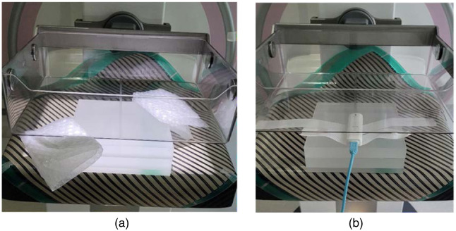
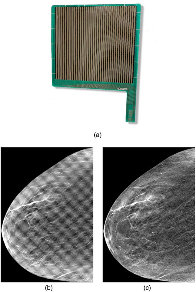
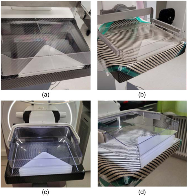
Approach: A radiation dose study was conducted on clinically available breast imaging systems with and without an MI sensor present. Our estimations were based on three approaches. In the first approach, exposure values were compared in paired clinical DBT and DBTMI acquisitions in 97 women. In the second approach polymethyl methacrylate (PMMA) phantoms of various thicknesses were used, and the average glandular dose (AGD) values were compared. Finally, a rectangular PMMA phantom with a 45 mm thickness was used, and the AGD values were estimated based on air kerma measurements with an electronic dosemeter.
Results: The relative increase in exposure estimated from digital imaging and communications in medicine headers when using an MI sensor in clinical DBTMI was . For the phantom measurements of various thicknesses of PMMA, the relative increases in the AGD for DM and DBT measurements were, on average, and , respectively. The relative increase in the AGD using the electronic dosemeter was in DM and in DBT. The average difference in dose between the methods was .
Conclusions: Our measurements suggest that the use of simultaneous breast radiography and MI increases the AGD by an average of . The increase in dose is within the acceptable values for mammography screening recommended by European guidelines.
期刊介绍:
JMI covers fundamental and translational research, as well as applications, focused on medical imaging, which continue to yield physical and biomedical advancements in the early detection, diagnostics, and therapy of disease as well as in the understanding of normal. The scope of JMI includes: Imaging physics, Tomographic reconstruction algorithms (such as those in CT and MRI), Image processing and deep learning, Computer-aided diagnosis and quantitative image analysis, Visualization and modeling, Picture archiving and communications systems (PACS), Image perception and observer performance, Technology assessment, Ultrasonic imaging, Image-guided procedures, Digital pathology, Biomedical applications of biomedical imaging. JMI allows for the peer-reviewed communication and archiving of scientific developments, translational and clinical applications, reviews, and recommendations for the field.


 分享
分享
 求助内容:
求助内容: 应助结果提醒方式:
 扫码关注我们
