Michael Aschner, Anatoly V. Skalny, Abel Santamaria, Joao B. T. Rocha, Borhan Mansouri, Yousef Tizabi, Roberto Madeddu, Rongzu Lu, Eunsook Lee, Alexey A. Tinkov
{"title":"铝诱导神经毒性和阿尔茨海默病的表观遗传学机制:聚焦非编码 RNA。","authors":"Michael Aschner, Anatoly V. Skalny, Abel Santamaria, Joao B. T. Rocha, Borhan Mansouri, Yousef Tizabi, Roberto Madeddu, Rongzu Lu, Eunsook Lee, Alexey A. Tinkov","doi":"10.1007/s11064-024-04214-9","DOIUrl":null,"url":null,"abstract":"<div><p>Aluminum (Al) is known to induce neurotoxic effects, potentially contributing to Alzheimer’s disease (AD) pathogenesis. Recent studies suggest that epigenetic modification may contribute to Al neurotoxicity, although the mechanisms are still debatable. Therefore, the objective of the present study was to summarize existing data on the involvement of epigenetic mechanisms in Al-induced neurotoxicity, especially AD-type pathology. Existing data demonstrate that Al exposure induces disruption in DNA methylation, histone modifications, and non-coding RNA expression in brains. Alterations in DNA methylation following Al exposure were shown to be mediated by changes in expression and activity of DNA methyltransferases (DNMTs) and ten-eleven translocation proteins (TETs). Al exposure was shown to reduce histone acetylation by up-regulating expression of histone deacetylases (HDACs) and impair histone methylation, ultimately contributing to down-regulation of brain-derived neurotrophic factor (BDNF) expression and activation of nuclear factor κB (NF-κB) signaling. Neurotoxic effects of Al exposure were also associated with aberrant expression of non-coding RNAs, especially microRNAs (miR). Al-induced patterns of miR expression were involved in development of AD-type pathology by increasing amyloid β (Aβ) production through up-regulation of Aβ precursor protein (APP) and β secretase (BACE1) expression (down-regulation of miR-29a/b, miR-101, miR-124, and Let-7c expression), increasing in neuroinflammation through NF-κB signaling (up-regulation of miR-9, miR-125b, miR-128, and 146a), as well as modulating other signaling pathways. Furthermore, reduced global DNA methylation, altered histone modification, and aberrant miRNA expression were associated with cognitive decline in Al-exposed subjects. However, further studies are required to evaluate the contribution of epigenetic mechanisms to Al-induced neurotoxicity and/or AD development.</p></div>","PeriodicalId":719,"journal":{"name":"Neurochemical Research","volume":"49 11","pages":"2988 - 3005"},"PeriodicalIF":3.8000,"publicationDate":"2024-07-27","publicationTypes":"Journal Article","fieldsOfStudy":null,"isOpenAccess":false,"openAccessPdf":"","citationCount":"0","resultStr":"{\"title\":\"Epigenetic Mechanisms of Aluminum-Induced Neurotoxicity and Alzheimer’s Disease: A Focus on Non-Coding RNAs\",\"authors\":\"Michael Aschner, Anatoly V. Skalny, Abel Santamaria, Joao B. T. Rocha, Borhan Mansouri, Yousef Tizabi, Roberto Madeddu, Rongzu Lu, Eunsook Lee, Alexey A. Tinkov\",\"doi\":\"10.1007/s11064-024-04214-9\",\"DOIUrl\":null,\"url\":null,\"abstract\":\"<div><p>Aluminum (Al) is known to induce neurotoxic effects, potentially contributing to Alzheimer’s disease (AD) pathogenesis. Recent studies suggest that epigenetic modification may contribute to Al neurotoxicity, although the mechanisms are still debatable. Therefore, the objective of the present study was to summarize existing data on the involvement of epigenetic mechanisms in Al-induced neurotoxicity, especially AD-type pathology. Existing data demonstrate that Al exposure induces disruption in DNA methylation, histone modifications, and non-coding RNA expression in brains. Alterations in DNA methylation following Al exposure were shown to be mediated by changes in expression and activity of DNA methyltransferases (DNMTs) and ten-eleven translocation proteins (TETs). Al exposure was shown to reduce histone acetylation by up-regulating expression of histone deacetylases (HDACs) and impair histone methylation, ultimately contributing to down-regulation of brain-derived neurotrophic factor (BDNF) expression and activation of nuclear factor κB (NF-κB) signaling. Neurotoxic effects of Al exposure were also associated with aberrant expression of non-coding RNAs, especially microRNAs (miR). Al-induced patterns of miR expression were involved in development of AD-type pathology by increasing amyloid β (Aβ) production through up-regulation of Aβ precursor protein (APP) and β secretase (BACE1) expression (down-regulation of miR-29a/b, miR-101, miR-124, and Let-7c expression), increasing in neuroinflammation through NF-κB signaling (up-regulation of miR-9, miR-125b, miR-128, and 146a), as well as modulating other signaling pathways. Furthermore, reduced global DNA methylation, altered histone modification, and aberrant miRNA expression were associated with cognitive decline in Al-exposed subjects. However, further studies are required to evaluate the contribution of epigenetic mechanisms to Al-induced neurotoxicity and/or AD development.</p></div>\",\"PeriodicalId\":719,\"journal\":{\"name\":\"Neurochemical Research\",\"volume\":\"49 11\",\"pages\":\"2988 - 3005\"},\"PeriodicalIF\":3.8000,\"publicationDate\":\"2024-07-27\",\"publicationTypes\":\"Journal Article\",\"fieldsOfStudy\":null,\"isOpenAccess\":false,\"openAccessPdf\":\"\",\"citationCount\":\"0\",\"resultStr\":null,\"platform\":\"Semanticscholar\",\"paperid\":null,\"PeriodicalName\":\"Neurochemical Research\",\"FirstCategoryId\":\"3\",\"ListUrlMain\":\"https://link.springer.com/article/10.1007/s11064-024-04214-9\",\"RegionNum\":3,\"RegionCategory\":\"医学\",\"ArticlePicture\":[],\"TitleCN\":null,\"AbstractTextCN\":null,\"PMCID\":null,\"EPubDate\":\"\",\"PubModel\":\"\",\"JCR\":\"Q2\",\"JCRName\":\"BIOCHEMISTRY & MOLECULAR BIOLOGY\",\"Score\":null,\"Total\":0}","platform":"Semanticscholar","paperid":null,"PeriodicalName":"Neurochemical Research","FirstCategoryId":"3","ListUrlMain":"https://link.springer.com/article/10.1007/s11064-024-04214-9","RegionNum":3,"RegionCategory":"医学","ArticlePicture":[],"TitleCN":null,"AbstractTextCN":null,"PMCID":null,"EPubDate":"","PubModel":"","JCR":"Q2","JCRName":"BIOCHEMISTRY & MOLECULAR BIOLOGY","Score":null,"Total":0}
引用次数: 0
摘要
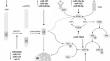
众所周知,铝(Al)具有神经毒性作用,可能导致阿尔茨海默病(AD)的发病。最近的研究表明,表观遗传修饰可能有助于铝的神经毒性,但其机制仍有待商榷。因此,本研究旨在总结表观遗传机制参与铝诱导的神经毒性,尤其是 AD 型病理学的现有数据。现有数据表明,铝暴露会诱导大脑中DNA甲基化、组蛋白修饰和非编码RNA表达的破坏。研究表明,暴露于铝后DNA甲基化的改变是由DNA甲基转移酶(DNMTs)和十-十一转位蛋白(TETs)的表达和活性变化介导的。研究表明,接触铝会通过上调组蛋白去乙酰化酶(HDACs)的表达来减少组蛋白乙酰化,并损害组蛋白甲基化,最终导致脑源性神经营养因子(BDNF)表达下调和核因子κB(NF-κB)信号的激活。铝暴露的神经毒性效应还与非编码 RNA,特别是微 RNA(miR)的异常表达有关。铝诱导的 miR 表达模式通过上调 Aβ 前体蛋白(APP)和 β 分泌酶(BACE1)的表达(下调 miR-29a/b、miR-101、miR-124 和 Let-7c 的表达),通过 NF-κB 信号转导增加神经炎症(上调 miR-9、miR-125b、miR-128 和 146a),以及调节其他信号通路。此外,全局 DNA 甲基化的减少、组蛋白修饰的改变和 miRNA 表达的异常与暴露于铝的受试者认知能力的下降有关。然而,要评估表观遗传机制对铝诱导的神经毒性和/或注意力缺失症发展的贡献,还需要进一步的研究。
Epigenetic Mechanisms of Aluminum-Induced Neurotoxicity and Alzheimer’s Disease: A Focus on Non-Coding RNAs
Aluminum (Al) is known to induce neurotoxic effects, potentially contributing to Alzheimer’s disease (AD) pathogenesis. Recent studies suggest that epigenetic modification may contribute to Al neurotoxicity, although the mechanisms are still debatable. Therefore, the objective of the present study was to summarize existing data on the involvement of epigenetic mechanisms in Al-induced neurotoxicity, especially AD-type pathology. Existing data demonstrate that Al exposure induces disruption in DNA methylation, histone modifications, and non-coding RNA expression in brains. Alterations in DNA methylation following Al exposure were shown to be mediated by changes in expression and activity of DNA methyltransferases (DNMTs) and ten-eleven translocation proteins (TETs). Al exposure was shown to reduce histone acetylation by up-regulating expression of histone deacetylases (HDACs) and impair histone methylation, ultimately contributing to down-regulation of brain-derived neurotrophic factor (BDNF) expression and activation of nuclear factor κB (NF-κB) signaling. Neurotoxic effects of Al exposure were also associated with aberrant expression of non-coding RNAs, especially microRNAs (miR). Al-induced patterns of miR expression were involved in development of AD-type pathology by increasing amyloid β (Aβ) production through up-regulation of Aβ precursor protein (APP) and β secretase (BACE1) expression (down-regulation of miR-29a/b, miR-101, miR-124, and Let-7c expression), increasing in neuroinflammation through NF-κB signaling (up-regulation of miR-9, miR-125b, miR-128, and 146a), as well as modulating other signaling pathways. Furthermore, reduced global DNA methylation, altered histone modification, and aberrant miRNA expression were associated with cognitive decline in Al-exposed subjects. However, further studies are required to evaluate the contribution of epigenetic mechanisms to Al-induced neurotoxicity and/or AD development.
期刊介绍:
Neurochemical Research is devoted to the rapid publication of studies that use neurochemical methodology in research on nervous system structure and function. The journal publishes original reports of experimental and clinical research results, perceptive reviews of significant problem areas in the neurosciences, brief comments of a methodological or interpretive nature, and research summaries conducted by leading scientists whose works are not readily available in English.


 分享
分享
 求助内容:
求助内容: 应助结果提醒方式:
 扫码关注我们
