Ibtisam Al-Musawi, Bethany H Dennis, Gavin J Clowry, Fiona E N LeBeau
{"title":"幼年α-突触核蛋白(A30P)转基因小鼠海马神经元兴奋性和神经炎症前驱变化的证据。","authors":"Ibtisam Al-Musawi, Bethany H Dennis, Gavin J Clowry, Fiona E N LeBeau","doi":"10.3389/frdem.2024.1404841","DOIUrl":null,"url":null,"abstract":"<p><strong>Introduction: </strong>Neuronal hyperexcitability and neuroinflammation are thought to occur at early stages in a range of neurodegenerative diseases. Neuroinflammation, notably activation of microglia, has been identified as a potential prodromal marker of dementia with Lewy bodies (DLB). Using a transgenic mouse model of DLB that over-expresses human mutant (A30P) alpha-synuclein (hα-syn) we have investigated whether early neuroinflammation is evident in the hippocampus in young pre-symptomatic animals.</p><p><strong>Methods: </strong>Previous studies have shown early hyperexcitability in the hippocampal CA3 region in male A30P mice at 2-4 months of age, therefore, in the current study we have immunostained this region for markers of neuronal activity (c-Fos), reactive astrocytes (glial fibrillary acidic protein, GFAP), microglia (ionizing calcium binding adapter protein 1, Iba-1) and reactive microglia (inducible nitric oxide synthase, iNOS).</p><p><strong>Results: </strong>We found an interesting biphasic change in the expression of c-Fos in A30P mice with high expression at 1 month, consistent with early onset of hyperexcitability, but lower expression from 2-4 months in male A30P mice compared to wild-type (WT) controls, possibly indicating chronic hyperexcitability. Neuroinflammation was indicated by significant increases in the % area of GFAP and the number of Iba-1+ cells that expressed iNOS immunoreactivity in the CA3 region in 2-4 months A30P male mice compared to WT controls. A similar increase in % area of GFAP was observed in female A30P mice, however, the Iba-1 count was not different between female WT and A30P mice. In WT mice aged 2-4 months only 4.6% of Iba-1+ cells co-expressed iNOS. In contrast, in age matched A30P mice 87% of cells co-expressed Iba-1 and iNOS. Although there was no difference in GFAP immunoreactivity at 1 month, Iba-1/iNOS co-expression was also increased in a cohort of 1 month old A30P mice.</p><p><strong>Discussion: </strong>Abnormal hα-syn expression in A30P mice caused early changes in network excitability, as indicated by c-Fos expression, and neuroinflammation which might contribute to disease progression.</p>","PeriodicalId":520000,"journal":{"name":"Frontiers in dementia","volume":"3 ","pages":"1404841"},"PeriodicalIF":0.0000,"publicationDate":"2024-06-17","publicationTypes":"Journal Article","fieldsOfStudy":null,"isOpenAccess":false,"openAccessPdf":"https://www.ncbi.nlm.nih.gov/pmc/articles/PMC11285622/pdf/","citationCount":"0","resultStr":"{\"title\":\"Evidence for prodromal changes in neuronal excitability and neuroinflammation in the hippocampus in young alpha-synuclein (A30P) transgenic mice.\",\"authors\":\"Ibtisam Al-Musawi, Bethany H Dennis, Gavin J Clowry, Fiona E N LeBeau\",\"doi\":\"10.3389/frdem.2024.1404841\",\"DOIUrl\":null,\"url\":null,\"abstract\":\"<p><strong>Introduction: </strong>Neuronal hyperexcitability and neuroinflammation are thought to occur at early stages in a range of neurodegenerative diseases. Neuroinflammation, notably activation of microglia, has been identified as a potential prodromal marker of dementia with Lewy bodies (DLB). Using a transgenic mouse model of DLB that over-expresses human mutant (A30P) alpha-synuclein (hα-syn) we have investigated whether early neuroinflammation is evident in the hippocampus in young pre-symptomatic animals.</p><p><strong>Methods: </strong>Previous studies have shown early hyperexcitability in the hippocampal CA3 region in male A30P mice at 2-4 months of age, therefore, in the current study we have immunostained this region for markers of neuronal activity (c-Fos), reactive astrocytes (glial fibrillary acidic protein, GFAP), microglia (ionizing calcium binding adapter protein 1, Iba-1) and reactive microglia (inducible nitric oxide synthase, iNOS).</p><p><strong>Results: </strong>We found an interesting biphasic change in the expression of c-Fos in A30P mice with high expression at 1 month, consistent with early onset of hyperexcitability, but lower expression from 2-4 months in male A30P mice compared to wild-type (WT) controls, possibly indicating chronic hyperexcitability. Neuroinflammation was indicated by significant increases in the % area of GFAP and the number of Iba-1+ cells that expressed iNOS immunoreactivity in the CA3 region in 2-4 months A30P male mice compared to WT controls. A similar increase in % area of GFAP was observed in female A30P mice, however, the Iba-1 count was not different between female WT and A30P mice. In WT mice aged 2-4 months only 4.6% of Iba-1+ cells co-expressed iNOS. In contrast, in age matched A30P mice 87% of cells co-expressed Iba-1 and iNOS. Although there was no difference in GFAP immunoreactivity at 1 month, Iba-1/iNOS co-expression was also increased in a cohort of 1 month old A30P mice.</p><p><strong>Discussion: </strong>Abnormal hα-syn expression in A30P mice caused early changes in network excitability, as indicated by c-Fos expression, and neuroinflammation which might contribute to disease progression.</p>\",\"PeriodicalId\":520000,\"journal\":{\"name\":\"Frontiers in dementia\",\"volume\":\"3 \",\"pages\":\"1404841\"},\"PeriodicalIF\":0.0000,\"publicationDate\":\"2024-06-17\",\"publicationTypes\":\"Journal Article\",\"fieldsOfStudy\":null,\"isOpenAccess\":false,\"openAccessPdf\":\"https://www.ncbi.nlm.nih.gov/pmc/articles/PMC11285622/pdf/\",\"citationCount\":\"0\",\"resultStr\":null,\"platform\":\"Semanticscholar\",\"paperid\":null,\"PeriodicalName\":\"Frontiers in dementia\",\"FirstCategoryId\":\"1085\",\"ListUrlMain\":\"https://doi.org/10.3389/frdem.2024.1404841\",\"RegionNum\":0,\"RegionCategory\":null,\"ArticlePicture\":[],\"TitleCN\":null,\"AbstractTextCN\":null,\"PMCID\":null,\"EPubDate\":\"2024/1/1 0:00:00\",\"PubModel\":\"eCollection\",\"JCR\":\"\",\"JCRName\":\"\",\"Score\":null,\"Total\":0}","platform":"Semanticscholar","paperid":null,"PeriodicalName":"Frontiers in dementia","FirstCategoryId":"1085","ListUrlMain":"https://doi.org/10.3389/frdem.2024.1404841","RegionNum":0,"RegionCategory":null,"ArticlePicture":[],"TitleCN":null,"AbstractTextCN":null,"PMCID":null,"EPubDate":"2024/1/1 0:00:00","PubModel":"eCollection","JCR":"","JCRName":"","Score":null,"Total":0}
Evidence for prodromal changes in neuronal excitability and neuroinflammation in the hippocampus in young alpha-synuclein (A30P) transgenic mice.
Introduction: Neuronal hyperexcitability and neuroinflammation are thought to occur at early stages in a range of neurodegenerative diseases. Neuroinflammation, notably activation of microglia, has been identified as a potential prodromal marker of dementia with Lewy bodies (DLB). Using a transgenic mouse model of DLB that over-expresses human mutant (A30P) alpha-synuclein (hα-syn) we have investigated whether early neuroinflammation is evident in the hippocampus in young pre-symptomatic animals.
Methods: Previous studies have shown early hyperexcitability in the hippocampal CA3 region in male A30P mice at 2-4 months of age, therefore, in the current study we have immunostained this region for markers of neuronal activity (c-Fos), reactive astrocytes (glial fibrillary acidic protein, GFAP), microglia (ionizing calcium binding adapter protein 1, Iba-1) and reactive microglia (inducible nitric oxide synthase, iNOS).
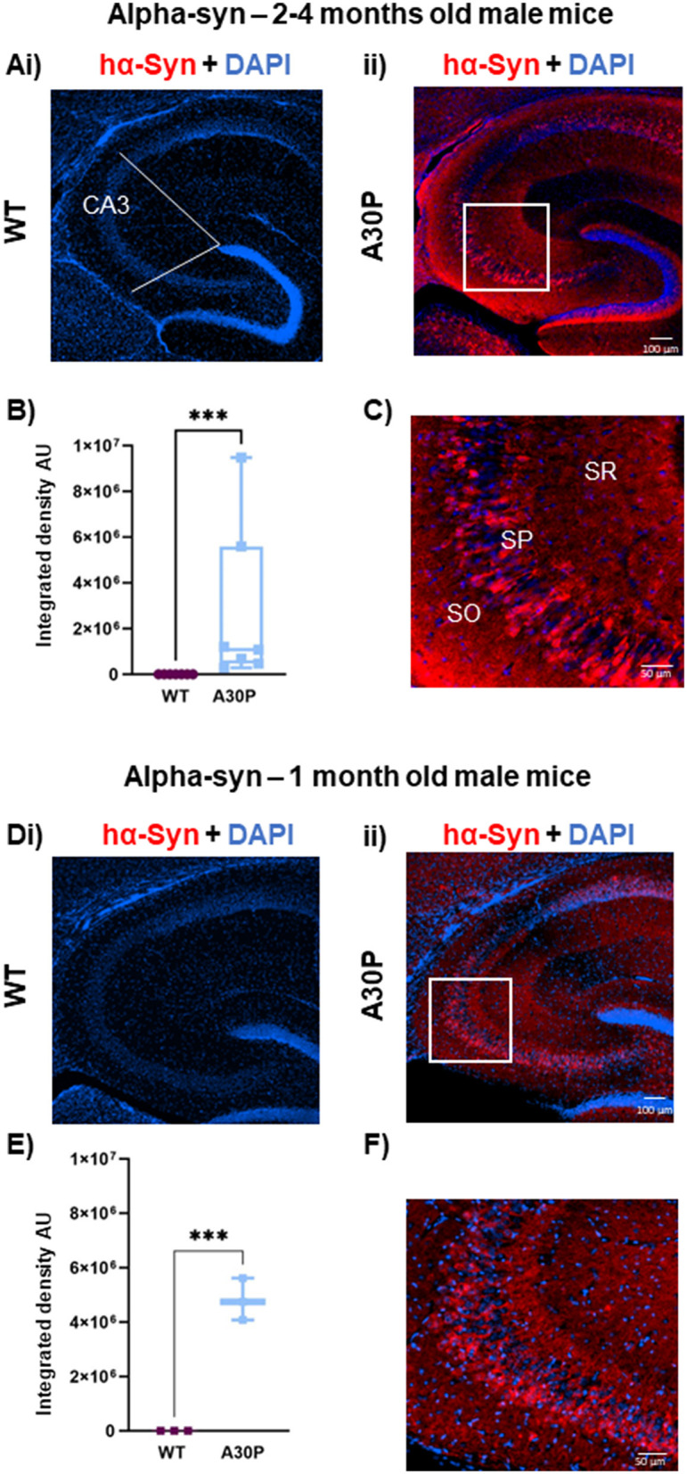
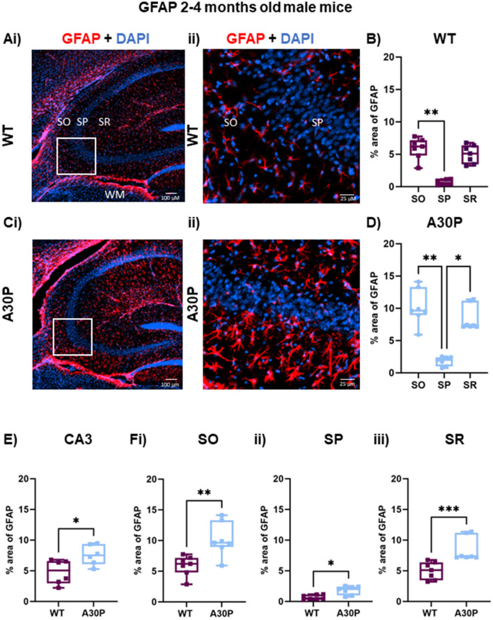
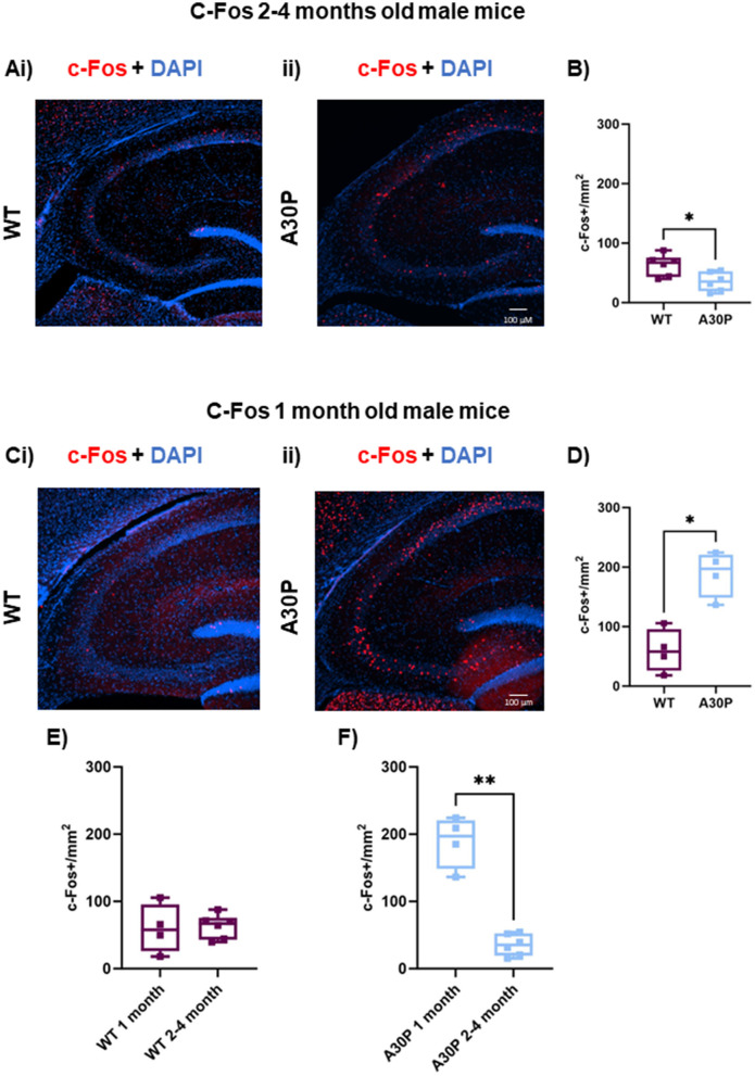
Results: We found an interesting biphasic change in the expression of c-Fos in A30P mice with high expression at 1 month, consistent with early onset of hyperexcitability, but lower expression from 2-4 months in male A30P mice compared to wild-type (WT) controls, possibly indicating chronic hyperexcitability. Neuroinflammation was indicated by significant increases in the % area of GFAP and the number of Iba-1+ cells that expressed iNOS immunoreactivity in the CA3 region in 2-4 months A30P male mice compared to WT controls. A similar increase in % area of GFAP was observed in female A30P mice, however, the Iba-1 count was not different between female WT and A30P mice. In WT mice aged 2-4 months only 4.6% of Iba-1+ cells co-expressed iNOS. In contrast, in age matched A30P mice 87% of cells co-expressed Iba-1 and iNOS. Although there was no difference in GFAP immunoreactivity at 1 month, Iba-1/iNOS co-expression was also increased in a cohort of 1 month old A30P mice.
Discussion: Abnormal hα-syn expression in A30P mice caused early changes in network excitability, as indicated by c-Fos expression, and neuroinflammation which might contribute to disease progression.


 分享
分享

 求助内容:
求助内容: 应助结果提醒方式:
 扫码关注我们
