Elia Halle, Tevel Amiel, Doron J Aframian, Tal Malik, Avital Rozenthal, Oren Shauly, Leo Joskowicz, Chen Nadler, Talia Yeshua
{"title":"虹膜锥束 CT 图像中腮腺导管闭合性涎腺的自动分割和深度学习分类。","authors":"Elia Halle, Tevel Amiel, Doron J Aframian, Tal Malik, Avital Rozenthal, Oren Shauly, Leo Joskowicz, Chen Nadler, Talia Yeshua","doi":"10.1007/s11548-024-03240-w","DOIUrl":null,"url":null,"abstract":"<p><strong>Purpose: </strong>This study addressed the challenge of detecting and classifying the severity of ductopenia in parotid glands, a structural abnormality characterized by a reduced number of salivary ducts, previously shown to be associated with salivary gland impairment. The aim of the study was to develop an automatic algorithm designed to improve diagnostic accuracy and efficiency in analyzing ductopenic parotid glands using sialo cone-beam CT (sialo-CBCT) images.</p><p><strong>Methods: </strong>We developed an end-to-end automatic pipeline consisting of three main steps: (1) region of interest (ROI) computation, (2) parotid gland segmentation using the Frangi filter, and (3) ductopenia case classification with a residual neural network (RNN) augmented by multidirectional maximum intensity projection (MIP) images. To explore the impact of the first two steps, the RNN was trained on three datasets: (1) original MIP images, (2) MIP images with predefined ROIs, and (3) MIP images after segmentation.</p><p><strong>Results: </strong>Evaluation was conducted on 126 parotid sialo-CBCT scans of normal, moderate, and severe ductopenic cases, yielding a high performance of 100% for the ROI computation and 89% for the gland segmentation. Improvements in accuracy and F1 score were noted among the original MIP images (accuracy: 0.73, F1 score: 0.53), ROI-predefined images (accuracy: 0.78, F1 score: 0.56), and segmented images (accuracy: 0.95, F1 score: 0.90). Notably, ductopenic detection sensitivity was 0.99 in the segmented dataset, highlighting the capabilities of the algorithm in detecting ductopenic cases.</p><p><strong>Conclusions: </strong>Our method, which combines classical image processing and deep learning techniques, offers a promising solution for automatic detection of parotid glands ductopenia in sialo-CBCT scans. This may be used for further research aimed at understanding the role of presence and severity of ductopenia in salivary gland dysfunction.</p>","PeriodicalId":51251,"journal":{"name":"International Journal of Computer Assisted Radiology and Surgery","volume":" ","pages":"21-30"},"PeriodicalIF":2.3000,"publicationDate":"2025-01-01","publicationTypes":"Journal Article","fieldsOfStudy":null,"isOpenAccess":false,"openAccessPdf":"","citationCount":"0","resultStr":"{\"title\":\"Automated segmentation and deep learning classification of ductopenic parotid salivary glands in sialo cone-beam CT images.\",\"authors\":\"Elia Halle, Tevel Amiel, Doron J Aframian, Tal Malik, Avital Rozenthal, Oren Shauly, Leo Joskowicz, Chen Nadler, Talia Yeshua\",\"doi\":\"10.1007/s11548-024-03240-w\",\"DOIUrl\":null,\"url\":null,\"abstract\":\"<p><strong>Purpose: </strong>This study addressed the challenge of detecting and classifying the severity of ductopenia in parotid glands, a structural abnormality characterized by a reduced number of salivary ducts, previously shown to be associated with salivary gland impairment. The aim of the study was to develop an automatic algorithm designed to improve diagnostic accuracy and efficiency in analyzing ductopenic parotid glands using sialo cone-beam CT (sialo-CBCT) images.</p><p><strong>Methods: </strong>We developed an end-to-end automatic pipeline consisting of three main steps: (1) region of interest (ROI) computation, (2) parotid gland segmentation using the Frangi filter, and (3) ductopenia case classification with a residual neural network (RNN) augmented by multidirectional maximum intensity projection (MIP) images. To explore the impact of the first two steps, the RNN was trained on three datasets: (1) original MIP images, (2) MIP images with predefined ROIs, and (3) MIP images after segmentation.</p><p><strong>Results: </strong>Evaluation was conducted on 126 parotid sialo-CBCT scans of normal, moderate, and severe ductopenic cases, yielding a high performance of 100% for the ROI computation and 89% for the gland segmentation. Improvements in accuracy and F1 score were noted among the original MIP images (accuracy: 0.73, F1 score: 0.53), ROI-predefined images (accuracy: 0.78, F1 score: 0.56), and segmented images (accuracy: 0.95, F1 score: 0.90). Notably, ductopenic detection sensitivity was 0.99 in the segmented dataset, highlighting the capabilities of the algorithm in detecting ductopenic cases.</p><p><strong>Conclusions: </strong>Our method, which combines classical image processing and deep learning techniques, offers a promising solution for automatic detection of parotid glands ductopenia in sialo-CBCT scans. This may be used for further research aimed at understanding the role of presence and severity of ductopenia in salivary gland dysfunction.</p>\",\"PeriodicalId\":51251,\"journal\":{\"name\":\"International Journal of Computer Assisted Radiology and Surgery\",\"volume\":\" \",\"pages\":\"21-30\"},\"PeriodicalIF\":2.3000,\"publicationDate\":\"2025-01-01\",\"publicationTypes\":\"Journal Article\",\"fieldsOfStudy\":null,\"isOpenAccess\":false,\"openAccessPdf\":\"\",\"citationCount\":\"0\",\"resultStr\":null,\"platform\":\"Semanticscholar\",\"paperid\":null,\"PeriodicalName\":\"International Journal of Computer Assisted Radiology and Surgery\",\"FirstCategoryId\":\"5\",\"ListUrlMain\":\"https://doi.org/10.1007/s11548-024-03240-w\",\"RegionNum\":3,\"RegionCategory\":\"医学\",\"ArticlePicture\":[],\"TitleCN\":null,\"AbstractTextCN\":null,\"PMCID\":null,\"EPubDate\":\"2024/7/31 0:00:00\",\"PubModel\":\"Epub\",\"JCR\":\"Q3\",\"JCRName\":\"ENGINEERING, BIOMEDICAL\",\"Score\":null,\"Total\":0}","platform":"Semanticscholar","paperid":null,"PeriodicalName":"International Journal of Computer Assisted Radiology and Surgery","FirstCategoryId":"5","ListUrlMain":"https://doi.org/10.1007/s11548-024-03240-w","RegionNum":3,"RegionCategory":"医学","ArticlePicture":[],"TitleCN":null,"AbstractTextCN":null,"PMCID":null,"EPubDate":"2024/7/31 0:00:00","PubModel":"Epub","JCR":"Q3","JCRName":"ENGINEERING, BIOMEDICAL","Score":null,"Total":0}
Automated segmentation and deep learning classification of ductopenic parotid salivary glands in sialo cone-beam CT images.
Purpose: This study addressed the challenge of detecting and classifying the severity of ductopenia in parotid glands, a structural abnormality characterized by a reduced number of salivary ducts, previously shown to be associated with salivary gland impairment. The aim of the study was to develop an automatic algorithm designed to improve diagnostic accuracy and efficiency in analyzing ductopenic parotid glands using sialo cone-beam CT (sialo-CBCT) images.
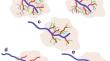
Methods: We developed an end-to-end automatic pipeline consisting of three main steps: (1) region of interest (ROI) computation, (2) parotid gland segmentation using the Frangi filter, and (3) ductopenia case classification with a residual neural network (RNN) augmented by multidirectional maximum intensity projection (MIP) images. To explore the impact of the first two steps, the RNN was trained on three datasets: (1) original MIP images, (2) MIP images with predefined ROIs, and (3) MIP images after segmentation.
Results: Evaluation was conducted on 126 parotid sialo-CBCT scans of normal, moderate, and severe ductopenic cases, yielding a high performance of 100% for the ROI computation and 89% for the gland segmentation. Improvements in accuracy and F1 score were noted among the original MIP images (accuracy: 0.73, F1 score: 0.53), ROI-predefined images (accuracy: 0.78, F1 score: 0.56), and segmented images (accuracy: 0.95, F1 score: 0.90). Notably, ductopenic detection sensitivity was 0.99 in the segmented dataset, highlighting the capabilities of the algorithm in detecting ductopenic cases.
Conclusions: Our method, which combines classical image processing and deep learning techniques, offers a promising solution for automatic detection of parotid glands ductopenia in sialo-CBCT scans. This may be used for further research aimed at understanding the role of presence and severity of ductopenia in salivary gland dysfunction.
期刊介绍:
The International Journal for Computer Assisted Radiology and Surgery (IJCARS) is a peer-reviewed journal that provides a platform for closing the gap between medical and technical disciplines, and encourages interdisciplinary research and development activities in an international environment.


 分享
分享
 求助内容:
求助内容: 应助结果提醒方式:
 扫码关注我们
