Kristina D Micheva, Jemima J Burden, Martina Schifferer
{"title":"阵列断层扫描:探索之路。","authors":"Kristina D Micheva, Jemima J Burden, Martina Schifferer","doi":"10.1515/mim-2024-0001","DOIUrl":null,"url":null,"abstract":"<p><p>Tissue slicing is at the core of many approaches to studying biological structures. Among the modern volume electron microscopy (vEM) methods, array tomography (AT) is based on serial ultramicrotomy, section collection onto solid support, imaging via light and/or scanning electron microscopy, and re-assembly of the serial images into a volume for analysis. While AT largely uses standard EM equipment, it provides several advantages, including long-term preservation of the sample and compatibility with multi-scale and multi-modal imaging. Furthermore, the collection of serial ultrathin sections improves axial resolution and provides access for molecular labeling, which is beneficial for light microscopy and immunolabeling, and facilitates correlation with EM. Despite these benefits, AT techniques are underrepresented in imaging facilities and labs, due to their perceived difficulty and lack of training opportunities. Here we point towards novel developments in serial sectioning and image analysis that facilitate the AT pipeline, and solutions to overcome constraints. Because no single vEM technique can serve all needs regarding field of view and resolution, we sketch a decision tree to aid researchers in navigating the plethora of options available. Lastly, we elaborate on the unexplored potential of AT approaches to add valuable insight in diverse biological fields.</p>","PeriodicalId":520012,"journal":{"name":"Methods in microscopy","volume":"1 1","pages":"9-17"},"PeriodicalIF":0.0000,"publicationDate":"2024-07-17","publicationTypes":"Journal Article","fieldsOfStudy":null,"isOpenAccess":false,"openAccessPdf":"https://www.ncbi.nlm.nih.gov/pmc/articles/PMC11308915/pdf/","citationCount":"0","resultStr":"{\"title\":\"Array tomography: trails to discovery.\",\"authors\":\"Kristina D Micheva, Jemima J Burden, Martina Schifferer\",\"doi\":\"10.1515/mim-2024-0001\",\"DOIUrl\":null,\"url\":null,\"abstract\":\"<p><p>Tissue slicing is at the core of many approaches to studying biological structures. Among the modern volume electron microscopy (vEM) methods, array tomography (AT) is based on serial ultramicrotomy, section collection onto solid support, imaging via light and/or scanning electron microscopy, and re-assembly of the serial images into a volume for analysis. While AT largely uses standard EM equipment, it provides several advantages, including long-term preservation of the sample and compatibility with multi-scale and multi-modal imaging. Furthermore, the collection of serial ultrathin sections improves axial resolution and provides access for molecular labeling, which is beneficial for light microscopy and immunolabeling, and facilitates correlation with EM. Despite these benefits, AT techniques are underrepresented in imaging facilities and labs, due to their perceived difficulty and lack of training opportunities. Here we point towards novel developments in serial sectioning and image analysis that facilitate the AT pipeline, and solutions to overcome constraints. Because no single vEM technique can serve all needs regarding field of view and resolution, we sketch a decision tree to aid researchers in navigating the plethora of options available. Lastly, we elaborate on the unexplored potential of AT approaches to add valuable insight in diverse biological fields.</p>\",\"PeriodicalId\":520012,\"journal\":{\"name\":\"Methods in microscopy\",\"volume\":\"1 1\",\"pages\":\"9-17\"},\"PeriodicalIF\":0.0000,\"publicationDate\":\"2024-07-17\",\"publicationTypes\":\"Journal Article\",\"fieldsOfStudy\":null,\"isOpenAccess\":false,\"openAccessPdf\":\"https://www.ncbi.nlm.nih.gov/pmc/articles/PMC11308915/pdf/\",\"citationCount\":\"0\",\"resultStr\":null,\"platform\":\"Semanticscholar\",\"paperid\":null,\"PeriodicalName\":\"Methods in microscopy\",\"FirstCategoryId\":\"1085\",\"ListUrlMain\":\"https://doi.org/10.1515/mim-2024-0001\",\"RegionNum\":0,\"RegionCategory\":null,\"ArticlePicture\":[],\"TitleCN\":null,\"AbstractTextCN\":null,\"PMCID\":null,\"EPubDate\":\"2024/4/1 0:00:00\",\"PubModel\":\"eCollection\",\"JCR\":\"\",\"JCRName\":\"\",\"Score\":null,\"Total\":0}","platform":"Semanticscholar","paperid":null,"PeriodicalName":"Methods in microscopy","FirstCategoryId":"1085","ListUrlMain":"https://doi.org/10.1515/mim-2024-0001","RegionNum":0,"RegionCategory":null,"ArticlePicture":[],"TitleCN":null,"AbstractTextCN":null,"PMCID":null,"EPubDate":"2024/4/1 0:00:00","PubModel":"eCollection","JCR":"","JCRName":"","Score":null,"Total":0}
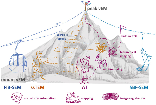
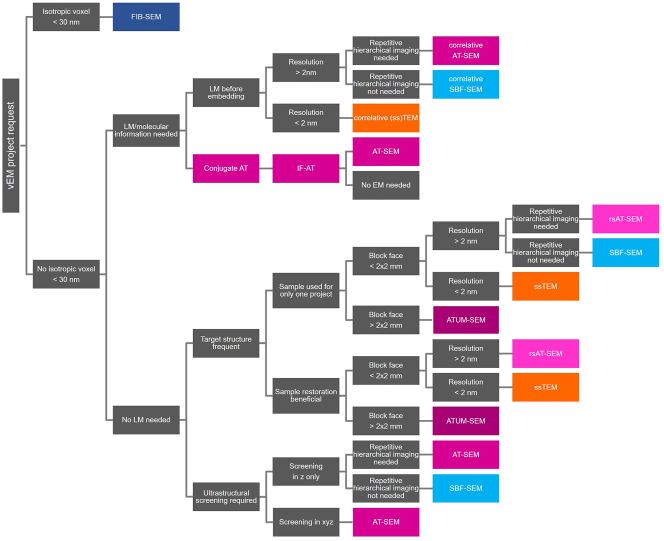
Tissue slicing is at the core of many approaches to studying biological structures. Among the modern volume electron microscopy (vEM) methods, array tomography (AT) is based on serial ultramicrotomy, section collection onto solid support, imaging via light and/or scanning electron microscopy, and re-assembly of the serial images into a volume for analysis. While AT largely uses standard EM equipment, it provides several advantages, including long-term preservation of the sample and compatibility with multi-scale and multi-modal imaging. Furthermore, the collection of serial ultrathin sections improves axial resolution and provides access for molecular labeling, which is beneficial for light microscopy and immunolabeling, and facilitates correlation with EM. Despite these benefits, AT techniques are underrepresented in imaging facilities and labs, due to their perceived difficulty and lack of training opportunities. Here we point towards novel developments in serial sectioning and image analysis that facilitate the AT pipeline, and solutions to overcome constraints. Because no single vEM technique can serve all needs regarding field of view and resolution, we sketch a decision tree to aid researchers in navigating the plethora of options available. Lastly, we elaborate on the unexplored potential of AT approaches to add valuable insight in diverse biological fields.


 分享
分享

 求助内容:
求助内容: 应助结果提醒方式:
 扫码关注我们
