Yi-Cheng Zhu, Li Zhou, Dao-Ming Zu, Shu-Hao Deng, Yuan Zhang, Jun Shan, Xiu-Rong Shi, Quan Jiang
{"title":"超级微血管成像和虚拟触摸成像量化在小儿肠系膜淋巴结炎诊断中的临床应用:有望提高诊断精确度的途径。","authors":"Yi-Cheng Zhu, Li Zhou, Dao-Ming Zu, Shu-Hao Deng, Yuan Zhang, Jun Shan, Xiu-Rong Shi, Quan Jiang","doi":"10.3233/CH-242305","DOIUrl":null,"url":null,"abstract":"<p><strong>Background: </strong>Mesenteric lymphadenitis (ML) demonstrates a distinctive inclination for the pediatric and adolescent demographic and the diagnosis of ML in young children poses a substantial challenge.</p><p><strong>Objective: </strong>This prospective study aimed to assess the diagnostic efficacy of Superb Microvascular Imaging (SMI) and Virtual Touch Tissue Imaging quantification (VTIQ) in distinguishing pediatric mesenteric lymphadentitis.</p><p><strong>Methods: </strong>We examined 82 mesentric lymph node (MLN) in pediatric patients with mesenteric lymphadentitis and 50 MLN in a healthy group. SMI was utilized to evaluate vascularity within the MLN, while MLN stiffness, quantified as shear wave velocity (SWV) in meters per second (m/s), was assessed using VTIQ. We compared the diagnostic performance of greyscale Ultrasound, US combined with SMI, US combined with VTIQ, and US combined with both SMI and VTIQ.</p><p><strong>Results: </strong>SMI revealed a significant distinction between mesenteric lymphadentitis and normal MLN (p < 0.001). MLN affected by mesenteric lymphadentis exhibited increased vascularity (marked vascularity: 13/82, 15.85%) compared to normal MLN (marked vascularity: 1/50, 2.00%). Statistically significant differences were observed in SWV values beween mesenteric lymphadentitis and normal MLN (all p-values <0.001). The mean and minimum SWV values for MLN with mesenteric lymphadentitis were 1.66±0.77 m/s and 1.51±0.53 m/s, respectively. Control group SWV values were approximately three times higher than those in the mesenteric lymphadenitis group. The highest area under the curve values were achieved with the combination of all three modalities (0.837, 95% confidence interval: 0.763- 0.896), followed by US + VTIQ (0.795, 0.716- 0.860), US + SMI (0.753, 0.670- 0.824) and US alone (0.642, 0.554- 0.724).</p><p><strong>Conclusion: </strong>SMI and VTIQ offer a promising noninvasive adjunct to grayscale ultrasound for identifying mesenteric lymphadentitis in pediatric patients.</p>","PeriodicalId":93943,"journal":{"name":"Clinical hemorheology and microcirculation","volume":" ","pages":"443-454"},"PeriodicalIF":0.0000,"publicationDate":"2024-01-01","publicationTypes":"Journal Article","fieldsOfStudy":null,"isOpenAccess":false,"openAccessPdf":"https://www.ncbi.nlm.nih.gov/pmc/articles/PMC11787815/pdf/","citationCount":"0","resultStr":"{\"title\":\"Clinical applications of superb microvascular imaging and virtual touch imaging quantification in pediatric mesenteric lymphadenitis diagnosis: A promising pathway to enhanced precision.\",\"authors\":\"Yi-Cheng Zhu, Li Zhou, Dao-Ming Zu, Shu-Hao Deng, Yuan Zhang, Jun Shan, Xiu-Rong Shi, Quan Jiang\",\"doi\":\"10.3233/CH-242305\",\"DOIUrl\":null,\"url\":null,\"abstract\":\"<p><strong>Background: </strong>Mesenteric lymphadenitis (ML) demonstrates a distinctive inclination for the pediatric and adolescent demographic and the diagnosis of ML in young children poses a substantial challenge.</p><p><strong>Objective: </strong>This prospective study aimed to assess the diagnostic efficacy of Superb Microvascular Imaging (SMI) and Virtual Touch Tissue Imaging quantification (VTIQ) in distinguishing pediatric mesenteric lymphadentitis.</p><p><strong>Methods: </strong>We examined 82 mesentric lymph node (MLN) in pediatric patients with mesenteric lymphadentitis and 50 MLN in a healthy group. SMI was utilized to evaluate vascularity within the MLN, while MLN stiffness, quantified as shear wave velocity (SWV) in meters per second (m/s), was assessed using VTIQ. We compared the diagnostic performance of greyscale Ultrasound, US combined with SMI, US combined with VTIQ, and US combined with both SMI and VTIQ.</p><p><strong>Results: </strong>SMI revealed a significant distinction between mesenteric lymphadentitis and normal MLN (p < 0.001). MLN affected by mesenteric lymphadentis exhibited increased vascularity (marked vascularity: 13/82, 15.85%) compared to normal MLN (marked vascularity: 1/50, 2.00%). Statistically significant differences were observed in SWV values beween mesenteric lymphadentitis and normal MLN (all p-values <0.001). The mean and minimum SWV values for MLN with mesenteric lymphadentitis were 1.66±0.77 m/s and 1.51±0.53 m/s, respectively. Control group SWV values were approximately three times higher than those in the mesenteric lymphadenitis group. The highest area under the curve values were achieved with the combination of all three modalities (0.837, 95% confidence interval: 0.763- 0.896), followed by US + VTIQ (0.795, 0.716- 0.860), US + SMI (0.753, 0.670- 0.824) and US alone (0.642, 0.554- 0.724).</p><p><strong>Conclusion: </strong>SMI and VTIQ offer a promising noninvasive adjunct to grayscale ultrasound for identifying mesenteric lymphadentitis in pediatric patients.</p>\",\"PeriodicalId\":93943,\"journal\":{\"name\":\"Clinical hemorheology and microcirculation\",\"volume\":\" \",\"pages\":\"443-454\"},\"PeriodicalIF\":0.0000,\"publicationDate\":\"2024-01-01\",\"publicationTypes\":\"Journal Article\",\"fieldsOfStudy\":null,\"isOpenAccess\":false,\"openAccessPdf\":\"https://www.ncbi.nlm.nih.gov/pmc/articles/PMC11787815/pdf/\",\"citationCount\":\"0\",\"resultStr\":null,\"platform\":\"Semanticscholar\",\"paperid\":null,\"PeriodicalName\":\"Clinical hemorheology and microcirculation\",\"FirstCategoryId\":\"1085\",\"ListUrlMain\":\"https://doi.org/10.3233/CH-242305\",\"RegionNum\":0,\"RegionCategory\":null,\"ArticlePicture\":[],\"TitleCN\":null,\"AbstractTextCN\":null,\"PMCID\":null,\"EPubDate\":\"\",\"PubModel\":\"\",\"JCR\":\"\",\"JCRName\":\"\",\"Score\":null,\"Total\":0}","platform":"Semanticscholar","paperid":null,"PeriodicalName":"Clinical hemorheology and microcirculation","FirstCategoryId":"1085","ListUrlMain":"https://doi.org/10.3233/CH-242305","RegionNum":0,"RegionCategory":null,"ArticlePicture":[],"TitleCN":null,"AbstractTextCN":null,"PMCID":null,"EPubDate":"","PubModel":"","JCR":"","JCRName":"","Score":null,"Total":0}
引用次数: 0
摘要
背景:肠系膜淋巴结炎(ML)在儿童和青少年人群中表现出明显的倾向性,幼儿肠系膜淋巴结炎的诊断是一个巨大的挑战:这项前瞻性研究旨在评估超级微血管成像(SMI)和虚拟触摸组织成像量化(VTIQ)在区分小儿肠系膜淋巴结炎方面的诊断效果:我们检查了82例小儿肠系膜淋巴结炎患者的肠系膜淋巴结(MLN)和50例健康组的肠系膜淋巴结。使用 SMI 评估 MLN 内的血管情况,同时使用 VTIQ 评估 MLN 的硬度,以剪切波速度(SWV)(米/秒)量化。我们比较了灰阶超声、US 结合 SMI、US 结合 VTIQ 以及 US 同时结合 SMI 和 VTIQ 的诊断性能:结果:SMI 显示肠系膜淋巴结炎与正常 MLN 有明显区别(P < 0.001)。与正常 MLN(明显血管:1/50,2.00%)相比,受肠系膜淋巴结炎影响的 MLN 表现出更高的血管性(明显血管:13/82,15.85%)。肠系膜淋巴结炎和正常 MLN 之间的 SWV 值差异具有统计学意义(所有 p 值均为结论值):SMI 和 VTIQ 可作为灰阶超声的无创辅助手段,用于鉴别儿科患者的肠系膜淋巴结炎。
Clinical applications of superb microvascular imaging and virtual touch imaging quantification in pediatric mesenteric lymphadenitis diagnosis: A promising pathway to enhanced precision.
Background: Mesenteric lymphadenitis (ML) demonstrates a distinctive inclination for the pediatric and adolescent demographic and the diagnosis of ML in young children poses a substantial challenge.
Objective: This prospective study aimed to assess the diagnostic efficacy of Superb Microvascular Imaging (SMI) and Virtual Touch Tissue Imaging quantification (VTIQ) in distinguishing pediatric mesenteric lymphadentitis.
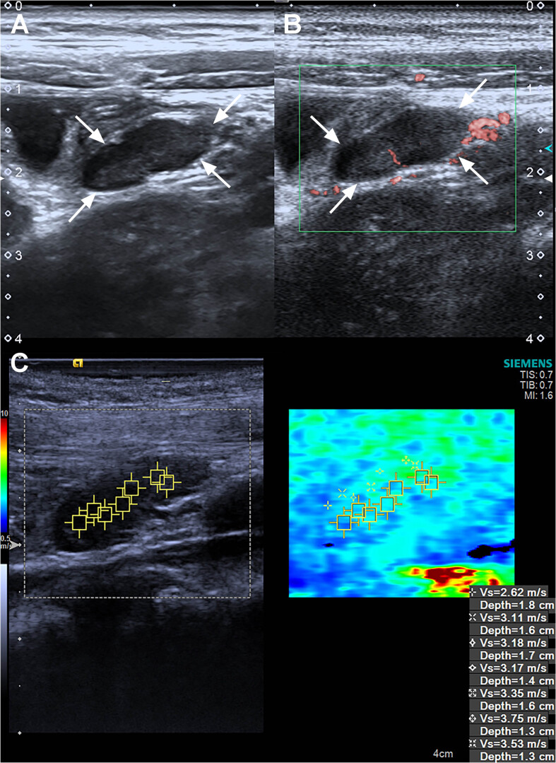
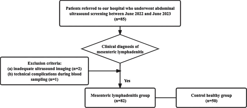
Methods: We examined 82 mesentric lymph node (MLN) in pediatric patients with mesenteric lymphadentitis and 50 MLN in a healthy group. SMI was utilized to evaluate vascularity within the MLN, while MLN stiffness, quantified as shear wave velocity (SWV) in meters per second (m/s), was assessed using VTIQ. We compared the diagnostic performance of greyscale Ultrasound, US combined with SMI, US combined with VTIQ, and US combined with both SMI and VTIQ.
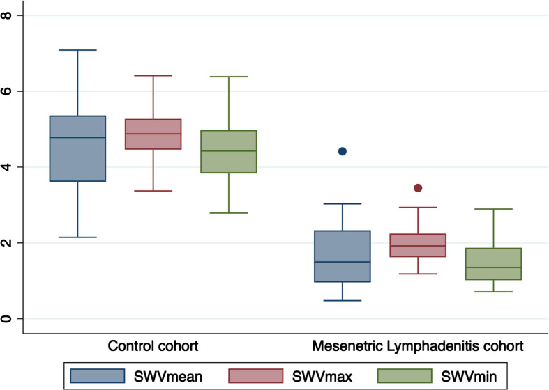
Results: SMI revealed a significant distinction between mesenteric lymphadentitis and normal MLN (p < 0.001). MLN affected by mesenteric lymphadentis exhibited increased vascularity (marked vascularity: 13/82, 15.85%) compared to normal MLN (marked vascularity: 1/50, 2.00%). Statistically significant differences were observed in SWV values beween mesenteric lymphadentitis and normal MLN (all p-values <0.001). The mean and minimum SWV values for MLN with mesenteric lymphadentitis were 1.66±0.77 m/s and 1.51±0.53 m/s, respectively. Control group SWV values were approximately three times higher than those in the mesenteric lymphadenitis group. The highest area under the curve values were achieved with the combination of all three modalities (0.837, 95% confidence interval: 0.763- 0.896), followed by US + VTIQ (0.795, 0.716- 0.860), US + SMI (0.753, 0.670- 0.824) and US alone (0.642, 0.554- 0.724).
Conclusion: SMI and VTIQ offer a promising noninvasive adjunct to grayscale ultrasound for identifying mesenteric lymphadentitis in pediatric patients.


 分享
分享

 求助内容:
求助内容: 应助结果提醒方式:
 扫码关注我们
