Fatih Alper, Adem Karaman, Ahmet Yalçın, Büşra Diyarbakır Şirinoğlu, Büşra Dinçer, Alptuğ Atila, Serhat Kaya, Taha Tavacı
{"title":"大鼠肾脏单剂量和多剂量注射钆沉积和 T1 信号强度变化的时间评估:一项实验研究","authors":"Fatih Alper, Adem Karaman, Ahmet Yalçın, Büşra Diyarbakır Şirinoğlu, Büşra Dinçer, Alptuğ Atila, Serhat Kaya, Taha Tavacı","doi":"10.5152/eurasianjmed.2024.23155","DOIUrl":null,"url":null,"abstract":"<p><strong>Background: </strong> Gadolinium deposition in biological tissues was first reported in patients with renal failure. We aimed to investigate gadolinium deposition in the rat kidney after exposure to single and multiple doses of gadolinium and evaluate deposition for 1- and 3-month periods. We also aimed to determine any correlation between the amount of deposition and T1-weighted image intensity.</p><p><strong>Methods: </strong> Seventy rats (5 animals per group) were included in the sample, and 9 groups received a single dose (0.3, 0.6, and 1.2 mmol/kg) of gadolinium, and 1 group for each dose was sacrificed at the end of the first day, week, and month. Four groups received weekly doses (0.3 and 0.6 mmol/kg) and were sacrificed at the end of 6 and 12 weeks. Measurement of T1 intensities was carried out with postinjection images before sacrifice, and deposition was determined using inductively coupled plasma mass spectrometry.</p><p><strong>Results: </strong> The number of injections was associated with increased gadolinium deposition (P <.001) in the kidney. After the weekly injections, the deposited gadolinium levels did not significantly difer between the low and medium doses at the end of the sixth week (P=.067). There was no agreement between the observers regarding the measurement of T1 signal intensity in both single-dose and multidose experiments (P=.263 and P=.307, respectively).</p><p><strong>Conclusion: </strong> Deposition was dose dependent in the postinjection stage in contrast to the late stage in which deposition was not associated with dose or number of injections until the 12th week. T1 signal intensity measurement is unreliable for assessing deposition in the rat kidney.</p>","PeriodicalId":53592,"journal":{"name":"Eurasian Journal of Medicine","volume":"56 1","pages":"47-51"},"PeriodicalIF":1.2000,"publicationDate":"2024-02-01","publicationTypes":"Journal Article","fieldsOfStudy":null,"isOpenAccess":false,"openAccessPdf":"https://www.ncbi.nlm.nih.gov/pmc/articles/PMC11059422/pdf/","citationCount":"0","resultStr":"{\"title\":\"Temporal Assessment of Gadolinium Deposition and T1 Signal Intensity Changes in Rat Kidney with Single and Multiple Doses of Injection: An Experimental Study.\",\"authors\":\"Fatih Alper, Adem Karaman, Ahmet Yalçın, Büşra Diyarbakır Şirinoğlu, Büşra Dinçer, Alptuğ Atila, Serhat Kaya, Taha Tavacı\",\"doi\":\"10.5152/eurasianjmed.2024.23155\",\"DOIUrl\":null,\"url\":null,\"abstract\":\"<p><strong>Background: </strong> Gadolinium deposition in biological tissues was first reported in patients with renal failure. We aimed to investigate gadolinium deposition in the rat kidney after exposure to single and multiple doses of gadolinium and evaluate deposition for 1- and 3-month periods. We also aimed to determine any correlation between the amount of deposition and T1-weighted image intensity.</p><p><strong>Methods: </strong> Seventy rats (5 animals per group) were included in the sample, and 9 groups received a single dose (0.3, 0.6, and 1.2 mmol/kg) of gadolinium, and 1 group for each dose was sacrificed at the end of the first day, week, and month. Four groups received weekly doses (0.3 and 0.6 mmol/kg) and were sacrificed at the end of 6 and 12 weeks. Measurement of T1 intensities was carried out with postinjection images before sacrifice, and deposition was determined using inductively coupled plasma mass spectrometry.</p><p><strong>Results: </strong> The number of injections was associated with increased gadolinium deposition (P <.001) in the kidney. After the weekly injections, the deposited gadolinium levels did not significantly difer between the low and medium doses at the end of the sixth week (P=.067). There was no agreement between the observers regarding the measurement of T1 signal intensity in both single-dose and multidose experiments (P=.263 and P=.307, respectively).</p><p><strong>Conclusion: </strong> Deposition was dose dependent in the postinjection stage in contrast to the late stage in which deposition was not associated with dose or number of injections until the 12th week. T1 signal intensity measurement is unreliable for assessing deposition in the rat kidney.</p>\",\"PeriodicalId\":53592,\"journal\":{\"name\":\"Eurasian Journal of Medicine\",\"volume\":\"56 1\",\"pages\":\"47-51\"},\"PeriodicalIF\":1.2000,\"publicationDate\":\"2024-02-01\",\"publicationTypes\":\"Journal Article\",\"fieldsOfStudy\":null,\"isOpenAccess\":false,\"openAccessPdf\":\"https://www.ncbi.nlm.nih.gov/pmc/articles/PMC11059422/pdf/\",\"citationCount\":\"0\",\"resultStr\":null,\"platform\":\"Semanticscholar\",\"paperid\":null,\"PeriodicalName\":\"Eurasian Journal of Medicine\",\"FirstCategoryId\":\"1085\",\"ListUrlMain\":\"https://doi.org/10.5152/eurasianjmed.2024.23155\",\"RegionNum\":0,\"RegionCategory\":null,\"ArticlePicture\":[],\"TitleCN\":null,\"AbstractTextCN\":null,\"PMCID\":null,\"EPubDate\":\"\",\"PubModel\":\"\",\"JCR\":\"Q3\",\"JCRName\":\"MEDICINE, GENERAL & INTERNAL\",\"Score\":null,\"Total\":0}","platform":"Semanticscholar","paperid":null,"PeriodicalName":"Eurasian Journal of Medicine","FirstCategoryId":"1085","ListUrlMain":"https://doi.org/10.5152/eurasianjmed.2024.23155","RegionNum":0,"RegionCategory":null,"ArticlePicture":[],"TitleCN":null,"AbstractTextCN":null,"PMCID":null,"EPubDate":"","PubModel":"","JCR":"Q3","JCRName":"MEDICINE, GENERAL & INTERNAL","Score":null,"Total":0}
Temporal Assessment of Gadolinium Deposition and T1 Signal Intensity Changes in Rat Kidney with Single and Multiple Doses of Injection: An Experimental Study.
Background: Gadolinium deposition in biological tissues was first reported in patients with renal failure. We aimed to investigate gadolinium deposition in the rat kidney after exposure to single and multiple doses of gadolinium and evaluate deposition for 1- and 3-month periods. We also aimed to determine any correlation between the amount of deposition and T1-weighted image intensity.
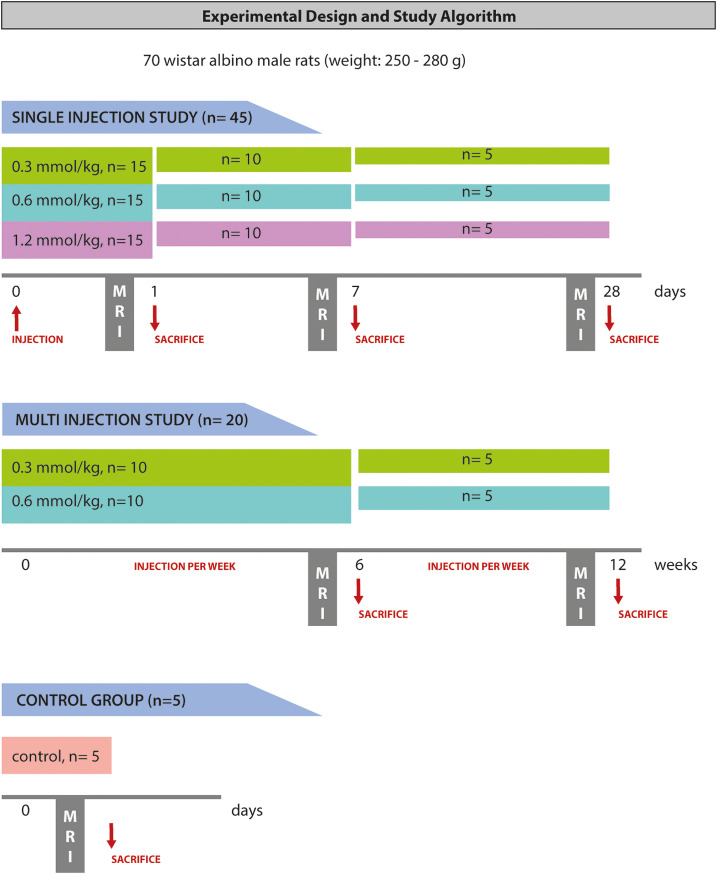
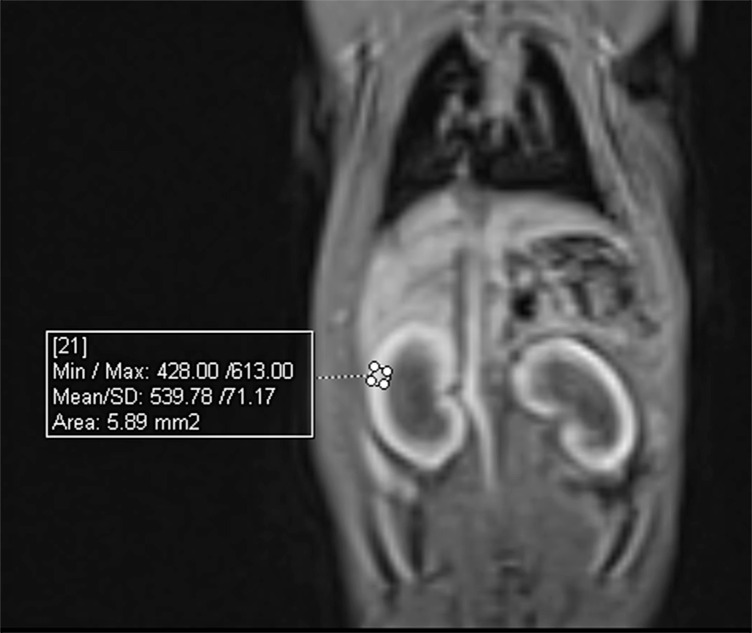
Methods: Seventy rats (5 animals per group) were included in the sample, and 9 groups received a single dose (0.3, 0.6, and 1.2 mmol/kg) of gadolinium, and 1 group for each dose was sacrificed at the end of the first day, week, and month. Four groups received weekly doses (0.3 and 0.6 mmol/kg) and were sacrificed at the end of 6 and 12 weeks. Measurement of T1 intensities was carried out with postinjection images before sacrifice, and deposition was determined using inductively coupled plasma mass spectrometry.
Results: The number of injections was associated with increased gadolinium deposition (P <.001) in the kidney. After the weekly injections, the deposited gadolinium levels did not significantly difer between the low and medium doses at the end of the sixth week (P=.067). There was no agreement between the observers regarding the measurement of T1 signal intensity in both single-dose and multidose experiments (P=.263 and P=.307, respectively).
Conclusion: Deposition was dose dependent in the postinjection stage in contrast to the late stage in which deposition was not associated with dose or number of injections until the 12th week. T1 signal intensity measurement is unreliable for assessing deposition in the rat kidney.
期刊介绍:
Eurasian Journal of Medicine (Eurasian J Med) is an international, scientific, open access periodical published by independent, unbiased, and triple-blinded peer-review principles. The journal is the official publication of Atatürk University School of Medicine and published triannually in February, June, and October. The publication language of the journal is English. The aim of the Eurasian Journal of Medicine is to publish original research papers of the highest scientific and clinical value in all medical fields. The Eurasian J Med also includes reviews, editorial short notes and letters to the editor that either as a comment related to recently published articles in our journal or as a case report. The target audience of the journal includes researchers, physicians and healthcare professionals who are interested or working in in all medical disciplines.


 分享
分享
 求助内容:
求助内容: 应助结果提醒方式:
 扫码关注我们
