{"title":"原发性胆汁性胆管炎患者的 CXCR6+CD8+T 细胞与临床病理参数之间的关系。","authors":"Huilian Shi, Xiangtao Xu, Shuangshuang Wang, Qinlei Chen, Fan Zhang, Haiyan Guo, Weiting Lu, Fei Qiao","doi":"10.1007/s12072-024-10715-0","DOIUrl":null,"url":null,"abstract":"<p><strong>Background: </strong>CXCR6+CD8+T cells have been implicated in the pathogenesis of various liver and autoimmune diseases. However, their involvement in primary biliary cholangitis (PBC) has not been elucidated.</p><p><strong>Methods: </strong>We used immunohistochemistry and flow cytometry to quantify CXCR6+CD8+T cells in hepatic tissue and peripheral blood samples obtained from CXCR6+CD8+T cells obtained from PBC patients. Then, we performed comprehensive statistical analyses to access the correlation between the abundance of these cells and clinical as well as pathological data across different stages of PBC.</p><p><strong>Results: </strong>Our research revealed that CXCR6+ cell frequencies in CD3+CD8+T cells from PBC patients significantly exceeded that of healthy controls (HCs) (2.24 vs. 0.61%, p < 0.01). A similar pattern emerged for hepatic CXCR6+CD8+T cell counts, which were notably higher in the PBC cohort compared to HCs. Our cohort consisted of 118 PBC patients, categorized into 62 early-stage (E-PBC) and 56 late-stage (L-PBC) cases. Notably, significant disparities existed between these groups in terms of liver enzyme and lipid profile levels (p < 0.05), with no notable differences observed in gender, age, blood counts, cholesterol levels, or autoantibodies (p > 0.05). Intriguingly, the quantity of hepatic CXCR6+CD8+T cells per high power field (HPF) was significantly elevated in both E-PBC and L-PBC patients as opposed to normal liver samples, indicating a substantial increase in these cells across all stages of PBC (p = 0.000). Spearman's rank correlation analysis showed a positive correlation between CXCR6+CD8+T cell counts and serum levels of Alkaline Phosphatase (AKP) and Gamma-Glutamyl Transferase (GGT), ANA, IgG and IgM, while revealing a negligible correlation with Alanine Aminotransferase (ALT) and Aspartate Aminotransferase (AST). Subsequent findings indicated significant variances in CXCR6+ cell numbers not only among different PBC stages but also across various degrees of inflammation and fibrosis (p ≤ 0.007). In a follow-up study post-Ursodeoxycholic Acid (UDCA) treatment, stark differences were identified in biochemical and immunohistochemical profiles between responder (31 patients) and non-responder (33 patients) groups (p < 0.05). A Wilcoxon rank-sum test further demonstrated a significant difference in the level of hepatic CXCR6+CD8+T cells between these two response groups (p = 0.002).</p><p><strong>Conclusion: </strong>CXCR6+CD8+T cells play a vital role in the pathogenesis of PBC, exhibiting correlations with the extent of inflammation, staging of liver fibrosis, and response to pharmacological interventions in PBC patients.</p>","PeriodicalId":12901,"journal":{"name":"Hepatology International","volume":" ","pages":"1555-1565"},"PeriodicalIF":6.1000,"publicationDate":"2024-10-01","publicationTypes":"Journal Article","fieldsOfStudy":null,"isOpenAccess":false,"openAccessPdf":"","citationCount":"0","resultStr":"{\"title\":\"The relationship between CXCR6+CD8+T cells and clinicopathological parameters in patients with primary biliary cholangitis.\",\"authors\":\"Huilian Shi, Xiangtao Xu, Shuangshuang Wang, Qinlei Chen, Fan Zhang, Haiyan Guo, Weiting Lu, Fei Qiao\",\"doi\":\"10.1007/s12072-024-10715-0\",\"DOIUrl\":null,\"url\":null,\"abstract\":\"<p><strong>Background: </strong>CXCR6+CD8+T cells have been implicated in the pathogenesis of various liver and autoimmune diseases. However, their involvement in primary biliary cholangitis (PBC) has not been elucidated.</p><p><strong>Methods: </strong>We used immunohistochemistry and flow cytometry to quantify CXCR6+CD8+T cells in hepatic tissue and peripheral blood samples obtained from CXCR6+CD8+T cells obtained from PBC patients. Then, we performed comprehensive statistical analyses to access the correlation between the abundance of these cells and clinical as well as pathological data across different stages of PBC.</p><p><strong>Results: </strong>Our research revealed that CXCR6+ cell frequencies in CD3+CD8+T cells from PBC patients significantly exceeded that of healthy controls (HCs) (2.24 vs. 0.61%, p < 0.01). A similar pattern emerged for hepatic CXCR6+CD8+T cell counts, which were notably higher in the PBC cohort compared to HCs. Our cohort consisted of 118 PBC patients, categorized into 62 early-stage (E-PBC) and 56 late-stage (L-PBC) cases. Notably, significant disparities existed between these groups in terms of liver enzyme and lipid profile levels (p < 0.05), with no notable differences observed in gender, age, blood counts, cholesterol levels, or autoantibodies (p > 0.05). Intriguingly, the quantity of hepatic CXCR6+CD8+T cells per high power field (HPF) was significantly elevated in both E-PBC and L-PBC patients as opposed to normal liver samples, indicating a substantial increase in these cells across all stages of PBC (p = 0.000). Spearman's rank correlation analysis showed a positive correlation between CXCR6+CD8+T cell counts and serum levels of Alkaline Phosphatase (AKP) and Gamma-Glutamyl Transferase (GGT), ANA, IgG and IgM, while revealing a negligible correlation with Alanine Aminotransferase (ALT) and Aspartate Aminotransferase (AST). Subsequent findings indicated significant variances in CXCR6+ cell numbers not only among different PBC stages but also across various degrees of inflammation and fibrosis (p ≤ 0.007). In a follow-up study post-Ursodeoxycholic Acid (UDCA) treatment, stark differences were identified in biochemical and immunohistochemical profiles between responder (31 patients) and non-responder (33 patients) groups (p < 0.05). A Wilcoxon rank-sum test further demonstrated a significant difference in the level of hepatic CXCR6+CD8+T cells between these two response groups (p = 0.002).</p><p><strong>Conclusion: </strong>CXCR6+CD8+T cells play a vital role in the pathogenesis of PBC, exhibiting correlations with the extent of inflammation, staging of liver fibrosis, and response to pharmacological interventions in PBC patients.</p>\",\"PeriodicalId\":12901,\"journal\":{\"name\":\"Hepatology International\",\"volume\":\" \",\"pages\":\"1555-1565\"},\"PeriodicalIF\":6.1000,\"publicationDate\":\"2024-10-01\",\"publicationTypes\":\"Journal Article\",\"fieldsOfStudy\":null,\"isOpenAccess\":false,\"openAccessPdf\":\"\",\"citationCount\":\"0\",\"resultStr\":null,\"platform\":\"Semanticscholar\",\"paperid\":null,\"PeriodicalName\":\"Hepatology International\",\"FirstCategoryId\":\"3\",\"ListUrlMain\":\"https://doi.org/10.1007/s12072-024-10715-0\",\"RegionNum\":2,\"RegionCategory\":\"医学\",\"ArticlePicture\":[],\"TitleCN\":null,\"AbstractTextCN\":null,\"PMCID\":null,\"EPubDate\":\"2024/8/12 0:00:00\",\"PubModel\":\"Epub\",\"JCR\":\"Q1\",\"JCRName\":\"GASTROENTEROLOGY & HEPATOLOGY\",\"Score\":null,\"Total\":0}","platform":"Semanticscholar","paperid":null,"PeriodicalName":"Hepatology International","FirstCategoryId":"3","ListUrlMain":"https://doi.org/10.1007/s12072-024-10715-0","RegionNum":2,"RegionCategory":"医学","ArticlePicture":[],"TitleCN":null,"AbstractTextCN":null,"PMCID":null,"EPubDate":"2024/8/12 0:00:00","PubModel":"Epub","JCR":"Q1","JCRName":"GASTROENTEROLOGY & HEPATOLOGY","Score":null,"Total":0}
The relationship between CXCR6+CD8+T cells and clinicopathological parameters in patients with primary biliary cholangitis.
Background: CXCR6+CD8+T cells have been implicated in the pathogenesis of various liver and autoimmune diseases. However, their involvement in primary biliary cholangitis (PBC) has not been elucidated.
Methods: We used immunohistochemistry and flow cytometry to quantify CXCR6+CD8+T cells in hepatic tissue and peripheral blood samples obtained from CXCR6+CD8+T cells obtained from PBC patients. Then, we performed comprehensive statistical analyses to access the correlation between the abundance of these cells and clinical as well as pathological data across different stages of PBC.
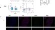
Results: Our research revealed that CXCR6+ cell frequencies in CD3+CD8+T cells from PBC patients significantly exceeded that of healthy controls (HCs) (2.24 vs. 0.61%, p < 0.01). A similar pattern emerged for hepatic CXCR6+CD8+T cell counts, which were notably higher in the PBC cohort compared to HCs. Our cohort consisted of 118 PBC patients, categorized into 62 early-stage (E-PBC) and 56 late-stage (L-PBC) cases. Notably, significant disparities existed between these groups in terms of liver enzyme and lipid profile levels (p < 0.05), with no notable differences observed in gender, age, blood counts, cholesterol levels, or autoantibodies (p > 0.05). Intriguingly, the quantity of hepatic CXCR6+CD8+T cells per high power field (HPF) was significantly elevated in both E-PBC and L-PBC patients as opposed to normal liver samples, indicating a substantial increase in these cells across all stages of PBC (p = 0.000). Spearman's rank correlation analysis showed a positive correlation between CXCR6+CD8+T cell counts and serum levels of Alkaline Phosphatase (AKP) and Gamma-Glutamyl Transferase (GGT), ANA, IgG and IgM, while revealing a negligible correlation with Alanine Aminotransferase (ALT) and Aspartate Aminotransferase (AST). Subsequent findings indicated significant variances in CXCR6+ cell numbers not only among different PBC stages but also across various degrees of inflammation and fibrosis (p ≤ 0.007). In a follow-up study post-Ursodeoxycholic Acid (UDCA) treatment, stark differences were identified in biochemical and immunohistochemical profiles between responder (31 patients) and non-responder (33 patients) groups (p < 0.05). A Wilcoxon rank-sum test further demonstrated a significant difference in the level of hepatic CXCR6+CD8+T cells between these two response groups (p = 0.002).
Conclusion: CXCR6+CD8+T cells play a vital role in the pathogenesis of PBC, exhibiting correlations with the extent of inflammation, staging of liver fibrosis, and response to pharmacological interventions in PBC patients.
期刊介绍:
Hepatology International is the official journal of the Asian Pacific Association for the Study of the Liver (APASL). This is a peer-reviewed journal featuring articles written by clinicians, clinical researchers and basic scientists is dedicated to research and patient care issues in hepatology. This journal will focus mainly on new and emerging technologies, cutting-edge science and advances in liver and biliary disorders.
Types of articles published:
-Original Research Articles related to clinical care and basic research
-Review Articles
-Consensus guidelines for diagnosis and treatment
-Clinical cases, images
-Selected Author Summaries
-Video Submissions


 分享
分享
 求助内容:
求助内容: 应助结果提醒方式:
 扫码关注我们
