Yi Yi, Lina Suo, Haixiu Ma, Ronghua Ma, Jing Zhao, Shaoqian Zhai, Haiyan Wang, Zhanhai Su
{"title":"MDM2 在血管生成中的作用:对内皮尖端细胞形成的影响。","authors":"Yi Yi, Lina Suo, Haixiu Ma, Ronghua Ma, Jing Zhao, Shaoqian Zhai, Haiyan Wang, Zhanhai Su","doi":"10.1007/s11626-024-00946-8","DOIUrl":null,"url":null,"abstract":"<p><p>In the present study, we examined the role of MDM2 in the angiogenesis process and its potential association with the sprouting of endothelial tip cells. To address this, we performed hypoxia-treated gastric cancer cells (HGC-27) to quantitative RT-PCR and Western blot analysis to measure the levels of MDM2 and VEGF-A mRNA and protein expression. Subsequently, we employed siRNA to disrupt MDM2 expression, followed by hypoxia treatment. The expression levels of MDM2 and VEGF-A mRNA and protein were subsequently reassessed. Additionally, ELISA was utilized to quantify the secretion levels of VEGF-A in each experimental group. A conditioned medium derived from HGC-27 cells treated with different agents was employed to assess its influence on the formation of EA.hy926 endothelial tip cells, using various techniques including Transwell plates migration assays, wound healing experiments, vascular formation assays, scanning electron microscopy, and immunofluorescence staining. These findings demonstrated that the in vitro knockdown of MDM2 in the conditioned medium exhibited significant inhibitory effects on endothelial cell migration, wound healing, and vascular formation. Additionally, the intervention led to a reduction in the presence of CD34<sup>+</sup> tip cells and the formation of filopodia in endothelial cells, while partially restoring the integrity of tight junctions. Subsequent examination utilizing RNA-seq revealed that the suppression of MDM2 in HGC-27 cells resulted in the downregulation of the PI3K/AKT signaling pathway. Consequently, this downregulation led to an elevation in angiogenic effects induced by hypoxia.</p>","PeriodicalId":13340,"journal":{"name":"In Vitro Cellular & Developmental Biology. Animal","volume":" ","pages":"983-995"},"PeriodicalIF":1.7000,"publicationDate":"2024-10-01","publicationTypes":"Journal Article","fieldsOfStudy":null,"isOpenAccess":false,"openAccessPdf":"","citationCount":"0","resultStr":"{\"title\":\"The role of MDM2 in angiogenesis: implications for endothelial tip cell formation.\",\"authors\":\"Yi Yi, Lina Suo, Haixiu Ma, Ronghua Ma, Jing Zhao, Shaoqian Zhai, Haiyan Wang, Zhanhai Su\",\"doi\":\"10.1007/s11626-024-00946-8\",\"DOIUrl\":null,\"url\":null,\"abstract\":\"<p><p>In the present study, we examined the role of MDM2 in the angiogenesis process and its potential association with the sprouting of endothelial tip cells. To address this, we performed hypoxia-treated gastric cancer cells (HGC-27) to quantitative RT-PCR and Western blot analysis to measure the levels of MDM2 and VEGF-A mRNA and protein expression. Subsequently, we employed siRNA to disrupt MDM2 expression, followed by hypoxia treatment. The expression levels of MDM2 and VEGF-A mRNA and protein were subsequently reassessed. Additionally, ELISA was utilized to quantify the secretion levels of VEGF-A in each experimental group. A conditioned medium derived from HGC-27 cells treated with different agents was employed to assess its influence on the formation of EA.hy926 endothelial tip cells, using various techniques including Transwell plates migration assays, wound healing experiments, vascular formation assays, scanning electron microscopy, and immunofluorescence staining. These findings demonstrated that the in vitro knockdown of MDM2 in the conditioned medium exhibited significant inhibitory effects on endothelial cell migration, wound healing, and vascular formation. Additionally, the intervention led to a reduction in the presence of CD34<sup>+</sup> tip cells and the formation of filopodia in endothelial cells, while partially restoring the integrity of tight junctions. Subsequent examination utilizing RNA-seq revealed that the suppression of MDM2 in HGC-27 cells resulted in the downregulation of the PI3K/AKT signaling pathway. Consequently, this downregulation led to an elevation in angiogenic effects induced by hypoxia.</p>\",\"PeriodicalId\":13340,\"journal\":{\"name\":\"In Vitro Cellular & Developmental Biology. Animal\",\"volume\":\" \",\"pages\":\"983-995\"},\"PeriodicalIF\":1.7000,\"publicationDate\":\"2024-10-01\",\"publicationTypes\":\"Journal Article\",\"fieldsOfStudy\":null,\"isOpenAccess\":false,\"openAccessPdf\":\"\",\"citationCount\":\"0\",\"resultStr\":null,\"platform\":\"Semanticscholar\",\"paperid\":null,\"PeriodicalName\":\"In Vitro Cellular & Developmental Biology. Animal\",\"FirstCategoryId\":\"99\",\"ListUrlMain\":\"https://doi.org/10.1007/s11626-024-00946-8\",\"RegionNum\":4,\"RegionCategory\":\"生物学\",\"ArticlePicture\":[],\"TitleCN\":null,\"AbstractTextCN\":null,\"PMCID\":null,\"EPubDate\":\"2024/8/12 0:00:00\",\"PubModel\":\"Epub\",\"JCR\":\"Q4\",\"JCRName\":\"CELL BIOLOGY\",\"Score\":null,\"Total\":0}","platform":"Semanticscholar","paperid":null,"PeriodicalName":"In Vitro Cellular & Developmental Biology. Animal","FirstCategoryId":"99","ListUrlMain":"https://doi.org/10.1007/s11626-024-00946-8","RegionNum":4,"RegionCategory":"生物学","ArticlePicture":[],"TitleCN":null,"AbstractTextCN":null,"PMCID":null,"EPubDate":"2024/8/12 0:00:00","PubModel":"Epub","JCR":"Q4","JCRName":"CELL BIOLOGY","Score":null,"Total":0}
The role of MDM2 in angiogenesis: implications for endothelial tip cell formation.
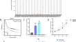
In the present study, we examined the role of MDM2 in the angiogenesis process and its potential association with the sprouting of endothelial tip cells. To address this, we performed hypoxia-treated gastric cancer cells (HGC-27) to quantitative RT-PCR and Western blot analysis to measure the levels of MDM2 and VEGF-A mRNA and protein expression. Subsequently, we employed siRNA to disrupt MDM2 expression, followed by hypoxia treatment. The expression levels of MDM2 and VEGF-A mRNA and protein were subsequently reassessed. Additionally, ELISA was utilized to quantify the secretion levels of VEGF-A in each experimental group. A conditioned medium derived from HGC-27 cells treated with different agents was employed to assess its influence on the formation of EA.hy926 endothelial tip cells, using various techniques including Transwell plates migration assays, wound healing experiments, vascular formation assays, scanning electron microscopy, and immunofluorescence staining. These findings demonstrated that the in vitro knockdown of MDM2 in the conditioned medium exhibited significant inhibitory effects on endothelial cell migration, wound healing, and vascular formation. Additionally, the intervention led to a reduction in the presence of CD34+ tip cells and the formation of filopodia in endothelial cells, while partially restoring the integrity of tight junctions. Subsequent examination utilizing RNA-seq revealed that the suppression of MDM2 in HGC-27 cells resulted in the downregulation of the PI3K/AKT signaling pathway. Consequently, this downregulation led to an elevation in angiogenic effects induced by hypoxia.
期刊介绍:
In Vitro Cellular & Developmental Biology - Animal is a journal of the Society for In Vitro Biology (SIVB). Original manuscripts reporting results of research in cellular, molecular, and developmental biology that employ or are relevant to organs, tissue, tumors, and cells in vitro will be considered for publication. Topics covered include:
Biotechnology;
Cell and Tissue Models;
Cell Growth/Differentiation/Apoptosis;
Cellular Pathology/Virology;
Cytokines/Growth Factors/Adhesion Factors;
Establishment of Cell Lines;
Signal Transduction;
Stem Cells;
Toxicology/Chemical Carcinogenesis;
Product Applications.


 分享
分享
 求助内容:
求助内容: 应助结果提醒方式:
 扫码关注我们
