Jing Shang Zhang, Jing Li, Jin Da Wang, Ying Xiong, Kai Cao, Meng Li, Kai Jie Wang, Ying Yan Mao, Jian Ying Liu, Xiu-Hua Wan
{"title":"小学生近视度数加深与视网膜厚度变化的关系","authors":"Jing Shang Zhang, Jing Li, Jin Da Wang, Ying Xiong, Kai Cao, Meng Li, Kai Jie Wang, Ying Yan Mao, Jian Ying Liu, Xiu-Hua Wan","doi":"10.1155/2024/1055700","DOIUrl":null,"url":null,"abstract":"<p><strong>Purpose: </strong>To observe the relationship between myopia progression and changes in retinal thickness during one year of follow-up among primary school children.</p><p><strong>Methods: </strong>The study included 1161 eyes of 708 myopic children, with 616 (53.06%) right eyes and 545 (46.94%) left eyes. The participants underwent a comprehensive ophthalmic examination, including visual acuity, axial length (AL), autorefraction, and optical coherence tomography (OCT) examination in 2016 and in 2017. An analysis was conducted on the differences in retinal thickness between different genders and between high myopia and nonhigh myopia. Furthermore, the study delved into the correlation between the progression of myopia and the changes of retinal thickness.</p><p><strong>Results: </strong>The average diopter was -1.83 ± 1.29D, average AL was 23.78 ± 0.94 mm, and average foveal thickness was 228.02 ± 23.00 <i>μ</i>m. For the inner retina, the median value [the lower quartile value, the upper quartile value] of the foveal thickness was thicker in the high myopia group than the nonhigh myopia group (67 [64; 74] <i>μ</i>m vs. 63 [56; 70] <i>μ</i>m), while the parafoveal region and perifoveal region were thinner in the high myopia group than the nonhigh myopia group (106 [100; 123] <i>μ</i>m vs. 124 [117; 130] <i>μ</i>m; 95.0 [93; 102] <i>μ</i>m vs. 104 [100; 108] <i>μ</i>m). Among all the children with myopia, 67.53% (784/1161) of them have a diopter progression within one year. The AL progression was 95.43% (1108/1161). The retinal thickness of all children has slightly increased in various regions. As the AL of the eye increased and the diopter decreased, the progression degree of inner retinal thickness and full retinal thickness (exclusive of full fovea) decreased.</p><p><strong>Conclusion: </strong>For the school-age myopic children, the inner foveal retinal thickness were thicker in highly myopic students than in the nonhighly myopic students, while the parafoveal and perifoveal retina were thinner in highly myopic students. The inner and full retinal thicknesses of male students were thicker than that of females. The progression of myopia mainly affected the changes of the inner retinal thickness in the one-year follow-up.</p>","PeriodicalId":16674,"journal":{"name":"Journal of Ophthalmology","volume":"2024 ","pages":"1055700"},"PeriodicalIF":1.9000,"publicationDate":"2024-08-06","publicationTypes":"Journal Article","fieldsOfStudy":null,"isOpenAccess":false,"openAccessPdf":"https://www.ncbi.nlm.nih.gov/pmc/articles/PMC11321887/pdf/","citationCount":"0","resultStr":"{\"title\":\"The Association of Myopia Progression with Changes in Retinal Thickness among Primary School Students with Myopia.\",\"authors\":\"Jing Shang Zhang, Jing Li, Jin Da Wang, Ying Xiong, Kai Cao, Meng Li, Kai Jie Wang, Ying Yan Mao, Jian Ying Liu, Xiu-Hua Wan\",\"doi\":\"10.1155/2024/1055700\",\"DOIUrl\":null,\"url\":null,\"abstract\":\"<p><strong>Purpose: </strong>To observe the relationship between myopia progression and changes in retinal thickness during one year of follow-up among primary school children.</p><p><strong>Methods: </strong>The study included 1161 eyes of 708 myopic children, with 616 (53.06%) right eyes and 545 (46.94%) left eyes. The participants underwent a comprehensive ophthalmic examination, including visual acuity, axial length (AL), autorefraction, and optical coherence tomography (OCT) examination in 2016 and in 2017. An analysis was conducted on the differences in retinal thickness between different genders and between high myopia and nonhigh myopia. Furthermore, the study delved into the correlation between the progression of myopia and the changes of retinal thickness.</p><p><strong>Results: </strong>The average diopter was -1.83 ± 1.29D, average AL was 23.78 ± 0.94 mm, and average foveal thickness was 228.02 ± 23.00 <i>μ</i>m. For the inner retina, the median value [the lower quartile value, the upper quartile value] of the foveal thickness was thicker in the high myopia group than the nonhigh myopia group (67 [64; 74] <i>μ</i>m vs. 63 [56; 70] <i>μ</i>m), while the parafoveal region and perifoveal region were thinner in the high myopia group than the nonhigh myopia group (106 [100; 123] <i>μ</i>m vs. 124 [117; 130] <i>μ</i>m; 95.0 [93; 102] <i>μ</i>m vs. 104 [100; 108] <i>μ</i>m). Among all the children with myopia, 67.53% (784/1161) of them have a diopter progression within one year. The AL progression was 95.43% (1108/1161). The retinal thickness of all children has slightly increased in various regions. As the AL of the eye increased and the diopter decreased, the progression degree of inner retinal thickness and full retinal thickness (exclusive of full fovea) decreased.</p><p><strong>Conclusion: </strong>For the school-age myopic children, the inner foveal retinal thickness were thicker in highly myopic students than in the nonhighly myopic students, while the parafoveal and perifoveal retina were thinner in highly myopic students. The inner and full retinal thicknesses of male students were thicker than that of females. The progression of myopia mainly affected the changes of the inner retinal thickness in the one-year follow-up.</p>\",\"PeriodicalId\":16674,\"journal\":{\"name\":\"Journal of Ophthalmology\",\"volume\":\"2024 \",\"pages\":\"1055700\"},\"PeriodicalIF\":1.9000,\"publicationDate\":\"2024-08-06\",\"publicationTypes\":\"Journal Article\",\"fieldsOfStudy\":null,\"isOpenAccess\":false,\"openAccessPdf\":\"https://www.ncbi.nlm.nih.gov/pmc/articles/PMC11321887/pdf/\",\"citationCount\":\"0\",\"resultStr\":null,\"platform\":\"Semanticscholar\",\"paperid\":null,\"PeriodicalName\":\"Journal of Ophthalmology\",\"FirstCategoryId\":\"3\",\"ListUrlMain\":\"https://doi.org/10.1155/2024/1055700\",\"RegionNum\":4,\"RegionCategory\":\"医学\",\"ArticlePicture\":[],\"TitleCN\":null,\"AbstractTextCN\":null,\"PMCID\":null,\"EPubDate\":\"2024/1/1 0:00:00\",\"PubModel\":\"eCollection\",\"JCR\":\"Q3\",\"JCRName\":\"OPHTHALMOLOGY\",\"Score\":null,\"Total\":0}","platform":"Semanticscholar","paperid":null,"PeriodicalName":"Journal of Ophthalmology","FirstCategoryId":"3","ListUrlMain":"https://doi.org/10.1155/2024/1055700","RegionNum":4,"RegionCategory":"医学","ArticlePicture":[],"TitleCN":null,"AbstractTextCN":null,"PMCID":null,"EPubDate":"2024/1/1 0:00:00","PubModel":"eCollection","JCR":"Q3","JCRName":"OPHTHALMOLOGY","Score":null,"Total":0}
The Association of Myopia Progression with Changes in Retinal Thickness among Primary School Students with Myopia.
Purpose: To observe the relationship between myopia progression and changes in retinal thickness during one year of follow-up among primary school children.
Methods: The study included 1161 eyes of 708 myopic children, with 616 (53.06%) right eyes and 545 (46.94%) left eyes. The participants underwent a comprehensive ophthalmic examination, including visual acuity, axial length (AL), autorefraction, and optical coherence tomography (OCT) examination in 2016 and in 2017. An analysis was conducted on the differences in retinal thickness between different genders and between high myopia and nonhigh myopia. Furthermore, the study delved into the correlation between the progression of myopia and the changes of retinal thickness.
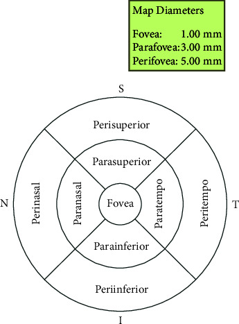
Results: The average diopter was -1.83 ± 1.29D, average AL was 23.78 ± 0.94 mm, and average foveal thickness was 228.02 ± 23.00 μm. For the inner retina, the median value [the lower quartile value, the upper quartile value] of the foveal thickness was thicker in the high myopia group than the nonhigh myopia group (67 [64; 74] μm vs. 63 [56; 70] μm), while the parafoveal region and perifoveal region were thinner in the high myopia group than the nonhigh myopia group (106 [100; 123] μm vs. 124 [117; 130] μm; 95.0 [93; 102] μm vs. 104 [100; 108] μm). Among all the children with myopia, 67.53% (784/1161) of them have a diopter progression within one year. The AL progression was 95.43% (1108/1161). The retinal thickness of all children has slightly increased in various regions. As the AL of the eye increased and the diopter decreased, the progression degree of inner retinal thickness and full retinal thickness (exclusive of full fovea) decreased.
Conclusion: For the school-age myopic children, the inner foveal retinal thickness were thicker in highly myopic students than in the nonhighly myopic students, while the parafoveal and perifoveal retina were thinner in highly myopic students. The inner and full retinal thicknesses of male students were thicker than that of females. The progression of myopia mainly affected the changes of the inner retinal thickness in the one-year follow-up.
期刊介绍:
Journal of Ophthalmology is a peer-reviewed, Open Access journal that publishes original research articles, review articles, and clinical studies related to the anatomy, physiology and diseases of the eye. Submissions should focus on new diagnostic and surgical techniques, instrument and therapy updates, as well as clinical trials and research findings.


 分享
分享
 求助内容:
求助内容: 应助结果提醒方式:
 扫码关注我们
