Guang-zhi Liao , Chun-hui He , Xin-qing Li , Yang Xiong , Li-yan Huang , An-ran Xin , Guo Ai , Man-qing Luo , Yu-hui Zhang , Jian Zhang
{"title":"探索心-脑和脑-心轴:大脑皮层结构与心血管疾病双向孟德尔随机研究的启示。","authors":"Guang-zhi Liao , Chun-hui He , Xin-qing Li , Yang Xiong , Li-yan Huang , An-ran Xin , Guo Ai , Man-qing Luo , Yu-hui Zhang , Jian Zhang","doi":"10.1016/j.nbd.2024.106636","DOIUrl":null,"url":null,"abstract":"<div><h3>Introduction</h3><p>The bidirectional relationship between the brain cortex and cardiovascular diseases (CVDs) remains inadequately explored.</p></div><div><h3>Methods</h3><p>This study used bidirectional Mendelian randomization (MR) analysis to explore the interactions between nine phenotypes associated with hypertension, heart failure, atrial fibrillation (AF), and coronary heart disease (CHD), and brain cortex measurements. These measurements included total surface area (SA), average thickness (TH), and the SA and TH of 34 regions defined by the Desikan-Killiany atlas. The nine traits were obtained from sources such as the UK Biobank and FinnGen, etc., while MRI-derived traits of cortical structure were sourced from the ENIGMA Consortium. The primary estimate was obtained using the inverse-variance weighted approach. A false discovery rate adjustment was applied to the p-values (resulting in q-values) in the analyses of regional cortical structures.</p></div><div><h3>Results</h3><p>A total of 1,260 two-sample MR analyses were conducted. Existing CHD demonstrated an influence on the SA of the banks of the superior temporal sulcus (bankssts) (q=0.018) and the superior frontal lobe (q=0.018), while hypertension was associated with changes in the TH of the lateral occipital region (q=0.02). Regarding the effects of the brain cortex on CVD incidence, total SA was significantly associated with the risk of CHD. Additionally, 16 and 3 regions exhibited significant effects on blood pressure and AF risk, respectively (q<0.05). These regions were primarily located in the frontal, temporal, and cingulate areas, which are associated with cognitive function and mood regulation.</p></div><div><h3>Conclusion</h3><p>The detection of cortical changes through MRI could aid in screening for potential neuropsychiatric disorders in individuals with established CVD. Moreover, abnormalities in cortical structure may predict future CVD risk, offering new insights for prevention and treatment strategies.</p></div>","PeriodicalId":19097,"journal":{"name":"Neurobiology of Disease","volume":"200 ","pages":"Article 106636"},"PeriodicalIF":5.6000,"publicationDate":"2024-10-01","publicationTypes":"Journal Article","fieldsOfStudy":null,"isOpenAccess":false,"openAccessPdf":"https://www.sciencedirect.com/science/article/pii/S0969996124002365/pdfft?md5=cd4d8aba5a6abc203083b2fa2efd01bc&pid=1-s2.0-S0969996124002365-main.pdf","citationCount":"0","resultStr":"{\"title\":\"Exploring the heart-brain and brain-heart axes: Insights from a bidirectional Mendelian randomization study on brain cortical structure and cardiovascular disease\",\"authors\":\"Guang-zhi Liao , Chun-hui He , Xin-qing Li , Yang Xiong , Li-yan Huang , An-ran Xin , Guo Ai , Man-qing Luo , Yu-hui Zhang , Jian Zhang\",\"doi\":\"10.1016/j.nbd.2024.106636\",\"DOIUrl\":null,\"url\":null,\"abstract\":\"<div><h3>Introduction</h3><p>The bidirectional relationship between the brain cortex and cardiovascular diseases (CVDs) remains inadequately explored.</p></div><div><h3>Methods</h3><p>This study used bidirectional Mendelian randomization (MR) analysis to explore the interactions between nine phenotypes associated with hypertension, heart failure, atrial fibrillation (AF), and coronary heart disease (CHD), and brain cortex measurements. These measurements included total surface area (SA), average thickness (TH), and the SA and TH of 34 regions defined by the Desikan-Killiany atlas. The nine traits were obtained from sources such as the UK Biobank and FinnGen, etc., while MRI-derived traits of cortical structure were sourced from the ENIGMA Consortium. The primary estimate was obtained using the inverse-variance weighted approach. A false discovery rate adjustment was applied to the p-values (resulting in q-values) in the analyses of regional cortical structures.</p></div><div><h3>Results</h3><p>A total of 1,260 two-sample MR analyses were conducted. Existing CHD demonstrated an influence on the SA of the banks of the superior temporal sulcus (bankssts) (q=0.018) and the superior frontal lobe (q=0.018), while hypertension was associated with changes in the TH of the lateral occipital region (q=0.02). Regarding the effects of the brain cortex on CVD incidence, total SA was significantly associated with the risk of CHD. Additionally, 16 and 3 regions exhibited significant effects on blood pressure and AF risk, respectively (q<0.05). These regions were primarily located in the frontal, temporal, and cingulate areas, which are associated with cognitive function and mood regulation.</p></div><div><h3>Conclusion</h3><p>The detection of cortical changes through MRI could aid in screening for potential neuropsychiatric disorders in individuals with established CVD. Moreover, abnormalities in cortical structure may predict future CVD risk, offering new insights for prevention and treatment strategies.</p></div>\",\"PeriodicalId\":19097,\"journal\":{\"name\":\"Neurobiology of Disease\",\"volume\":\"200 \",\"pages\":\"Article 106636\"},\"PeriodicalIF\":5.6000,\"publicationDate\":\"2024-10-01\",\"publicationTypes\":\"Journal Article\",\"fieldsOfStudy\":null,\"isOpenAccess\":false,\"openAccessPdf\":\"https://www.sciencedirect.com/science/article/pii/S0969996124002365/pdfft?md5=cd4d8aba5a6abc203083b2fa2efd01bc&pid=1-s2.0-S0969996124002365-main.pdf\",\"citationCount\":\"0\",\"resultStr\":null,\"platform\":\"Semanticscholar\",\"paperid\":null,\"PeriodicalName\":\"Neurobiology of Disease\",\"FirstCategoryId\":\"3\",\"ListUrlMain\":\"https://www.sciencedirect.com/science/article/pii/S0969996124002365\",\"RegionNum\":2,\"RegionCategory\":\"医学\",\"ArticlePicture\":[],\"TitleCN\":null,\"AbstractTextCN\":null,\"PMCID\":null,\"EPubDate\":\"2024/8/12 0:00:00\",\"PubModel\":\"Epub\",\"JCR\":\"Q1\",\"JCRName\":\"NEUROSCIENCES\",\"Score\":null,\"Total\":0}","platform":"Semanticscholar","paperid":null,"PeriodicalName":"Neurobiology of Disease","FirstCategoryId":"3","ListUrlMain":"https://www.sciencedirect.com/science/article/pii/S0969996124002365","RegionNum":2,"RegionCategory":"医学","ArticlePicture":[],"TitleCN":null,"AbstractTextCN":null,"PMCID":null,"EPubDate":"2024/8/12 0:00:00","PubModel":"Epub","JCR":"Q1","JCRName":"NEUROSCIENCES","Score":null,"Total":0}
Exploring the heart-brain and brain-heart axes: Insights from a bidirectional Mendelian randomization study on brain cortical structure and cardiovascular disease
Introduction
The bidirectional relationship between the brain cortex and cardiovascular diseases (CVDs) remains inadequately explored.
Methods
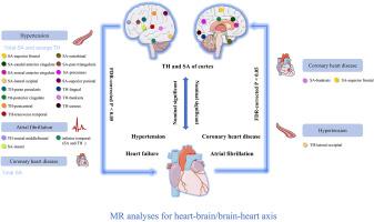
This study used bidirectional Mendelian randomization (MR) analysis to explore the interactions between nine phenotypes associated with hypertension, heart failure, atrial fibrillation (AF), and coronary heart disease (CHD), and brain cortex measurements. These measurements included total surface area (SA), average thickness (TH), and the SA and TH of 34 regions defined by the Desikan-Killiany atlas. The nine traits were obtained from sources such as the UK Biobank and FinnGen, etc., while MRI-derived traits of cortical structure were sourced from the ENIGMA Consortium. The primary estimate was obtained using the inverse-variance weighted approach. A false discovery rate adjustment was applied to the p-values (resulting in q-values) in the analyses of regional cortical structures.
Results
A total of 1,260 two-sample MR analyses were conducted. Existing CHD demonstrated an influence on the SA of the banks of the superior temporal sulcus (bankssts) (q=0.018) and the superior frontal lobe (q=0.018), while hypertension was associated with changes in the TH of the lateral occipital region (q=0.02). Regarding the effects of the brain cortex on CVD incidence, total SA was significantly associated with the risk of CHD. Additionally, 16 and 3 regions exhibited significant effects on blood pressure and AF risk, respectively (q<0.05). These regions were primarily located in the frontal, temporal, and cingulate areas, which are associated with cognitive function and mood regulation.
Conclusion
The detection of cortical changes through MRI could aid in screening for potential neuropsychiatric disorders in individuals with established CVD. Moreover, abnormalities in cortical structure may predict future CVD risk, offering new insights for prevention and treatment strategies.
期刊介绍:
Neurobiology of Disease is a major international journal at the interface between basic and clinical neuroscience. The journal provides a forum for the publication of top quality research papers on: molecular and cellular definitions of disease mechanisms, the neural systems and underpinning behavioral disorders, the genetics of inherited neurological and psychiatric diseases, nervous system aging, and findings relevant to the development of new therapies.


 分享
分享
 求助内容:
求助内容: 应助结果提醒方式:
 扫码关注我们
