Nancy Castro Borjas, Max Anstötz, Gianmaria Maccaferri
{"title":"小鼠泡状锥体神经元接收 GABA 能输入的细胞类型特异性受多层多样性的制约。","authors":"Nancy Castro Borjas, Max Anstötz, Gianmaria Maccaferri","doi":"10.1113/JP286679","DOIUrl":null,"url":null,"abstract":"<div>\n \n <section>\n \n \n <div>The subiculum is a key region of the brain involved in the initiation of pathological activity in temporal lobe epilepsy, and local GABAergic inhibition is essential to prevent subicular-originated epileptiform discharges. Subicular pyramidal cells may be easily distinguished into two classes based on their different firing patterns. Here, we have compared the strength of the GABAa receptor-mediated inhibitory postsynaptic currents received by regular- <i>vs</i>. burst-firing subicular neurons and their dynamic modulation by the activation of μ opioid receptors. We have taken advantage of the sequential re-patching of the same cell to initially classify pyramidal neurons according to their firing patters, and then to measure GABAergic events triggered by the optogenetic stimulation of parvalbumin- and somatostatin-expressing interneurons. Activation of parvalbumin-expressing cells generated larger responses in postsynaptic burst-firing neurons whereas the opposite was observed for currents evoked by the stimulation of somatostatin-expressing interneurons. In all cases, events depended critically on ω-agatoxin IVA- but not on ω-conotoxin GVIA-sensitive calcium channels. Optogenetic GABAergic input originating from both parvalbumin- and somatostatin-expressing cells was reduced in amplitude following the exposure to a μ opioid receptor agonist. The kinetics of this pharmacological sensitivity was different in regular- <i>vs</i>. burst-firing neurons, but only when responses were evoked by the activation of parvalbumin-expressing neurons, whereas no differences were observed when somatostatin-expressing cells were stimulated. In conclusion, our results show that a high degree of complexity regulates the organizing principles of subicular GABAergic inhibition, with the interaction of pre- and postsynaptic diversity at multiple levels.\n\n <figure>\n <div><picture>\n <source></source></picture><p></p>\n </div>\n </figure>\n </div>\n </section>\n \n <section>\n \n <h3> Key points</h3>\n \n <div>\n <ul>\n \n <li>Optogenetic stimulation of parvalbumin- and somatostatin-expressing interneurons (PVs and SOMs) triggers inhibitory postsynaptic currents (IPSCs) in both regular- and burst-firing (RFs and BFs) subicular pyramidal cells.</li>\n \n <li>The amplitude of optogenetically evoked IPSCs from PVs (PV-opto IPSCs) is larger in BFs whereas IPSCs generated by the light activation of SOMs (SOM-opto IPSCs) are larger in RFs.</li>\n \n <li>Both PV- and SOM-opto IPSCs critically depend on ω-agatoxin IVA-sensitive P/Q type voltage-gated calcium channels, whereas no major effects are observed following exposure to ω-conotoxin GVIA, suggesting no significant involvement of N-type channels.</li>\n \n <li>The amplitude of both PV- and SOM-opto IPSCs is reduced by the probable pharmacological activation of presynaptic μ opioid receptors, with a faster kinetics of the effect observed in PV-opto IPSCs from RFs <i>vs</i>. BFs, but not in SOM-opto IPSCs.</li>\n \n <li>These results help us understand the complex interactions between different layers of diversity regulating GABAergic input onto subicular microcircuits.</li>\n </ul>\n </div>\n </section>\n </div>","PeriodicalId":50088,"journal":{"name":"Journal of Physiology-London","volume":null,"pages":null},"PeriodicalIF":4.7000,"publicationDate":"2024-08-14","publicationTypes":"Journal Article","fieldsOfStudy":null,"isOpenAccess":false,"openAccessPdf":"https://onlinelibrary.wiley.com/doi/epdf/10.1113/JP286679","citationCount":"0","resultStr":"{\"title\":\"Multiple layers of diversity govern the cell type specificity of GABAergic input received by mouse subicular pyramidal neurons\",\"authors\":\"Nancy Castro Borjas, Max Anstötz, Gianmaria Maccaferri\",\"doi\":\"10.1113/JP286679\",\"DOIUrl\":null,\"url\":null,\"abstract\":\"<div>\\n \\n <section>\\n \\n \\n <div>The subiculum is a key region of the brain involved in the initiation of pathological activity in temporal lobe epilepsy, and local GABAergic inhibition is essential to prevent subicular-originated epileptiform discharges. Subicular pyramidal cells may be easily distinguished into two classes based on their different firing patterns. Here, we have compared the strength of the GABAa receptor-mediated inhibitory postsynaptic currents received by regular- <i>vs</i>. burst-firing subicular neurons and their dynamic modulation by the activation of μ opioid receptors. We have taken advantage of the sequential re-patching of the same cell to initially classify pyramidal neurons according to their firing patters, and then to measure GABAergic events triggered by the optogenetic stimulation of parvalbumin- and somatostatin-expressing interneurons. Activation of parvalbumin-expressing cells generated larger responses in postsynaptic burst-firing neurons whereas the opposite was observed for currents evoked by the stimulation of somatostatin-expressing interneurons. In all cases, events depended critically on ω-agatoxin IVA- but not on ω-conotoxin GVIA-sensitive calcium channels. Optogenetic GABAergic input originating from both parvalbumin- and somatostatin-expressing cells was reduced in amplitude following the exposure to a μ opioid receptor agonist. The kinetics of this pharmacological sensitivity was different in regular- <i>vs</i>. burst-firing neurons, but only when responses were evoked by the activation of parvalbumin-expressing neurons, whereas no differences were observed when somatostatin-expressing cells were stimulated. In conclusion, our results show that a high degree of complexity regulates the organizing principles of subicular GABAergic inhibition, with the interaction of pre- and postsynaptic diversity at multiple levels.\\n\\n <figure>\\n <div><picture>\\n <source></source></picture><p></p>\\n </div>\\n </figure>\\n </div>\\n </section>\\n \\n <section>\\n \\n <h3> Key points</h3>\\n \\n <div>\\n <ul>\\n \\n <li>Optogenetic stimulation of parvalbumin- and somatostatin-expressing interneurons (PVs and SOMs) triggers inhibitory postsynaptic currents (IPSCs) in both regular- and burst-firing (RFs and BFs) subicular pyramidal cells.</li>\\n \\n <li>The amplitude of optogenetically evoked IPSCs from PVs (PV-opto IPSCs) is larger in BFs whereas IPSCs generated by the light activation of SOMs (SOM-opto IPSCs) are larger in RFs.</li>\\n \\n <li>Both PV- and SOM-opto IPSCs critically depend on ω-agatoxin IVA-sensitive P/Q type voltage-gated calcium channels, whereas no major effects are observed following exposure to ω-conotoxin GVIA, suggesting no significant involvement of N-type channels.</li>\\n \\n <li>The amplitude of both PV- and SOM-opto IPSCs is reduced by the probable pharmacological activation of presynaptic μ opioid receptors, with a faster kinetics of the effect observed in PV-opto IPSCs from RFs <i>vs</i>. BFs, but not in SOM-opto IPSCs.</li>\\n \\n <li>These results help us understand the complex interactions between different layers of diversity regulating GABAergic input onto subicular microcircuits.</li>\\n </ul>\\n </div>\\n </section>\\n </div>\",\"PeriodicalId\":50088,\"journal\":{\"name\":\"Journal of Physiology-London\",\"volume\":null,\"pages\":null},\"PeriodicalIF\":4.7000,\"publicationDate\":\"2024-08-14\",\"publicationTypes\":\"Journal Article\",\"fieldsOfStudy\":null,\"isOpenAccess\":false,\"openAccessPdf\":\"https://onlinelibrary.wiley.com/doi/epdf/10.1113/JP286679\",\"citationCount\":\"0\",\"resultStr\":null,\"platform\":\"Semanticscholar\",\"paperid\":null,\"PeriodicalName\":\"Journal of Physiology-London\",\"FirstCategoryId\":\"3\",\"ListUrlMain\":\"https://onlinelibrary.wiley.com/doi/10.1113/JP286679\",\"RegionNum\":2,\"RegionCategory\":\"医学\",\"ArticlePicture\":[],\"TitleCN\":null,\"AbstractTextCN\":null,\"PMCID\":null,\"EPubDate\":\"\",\"PubModel\":\"\",\"JCR\":\"Q1\",\"JCRName\":\"NEUROSCIENCES\",\"Score\":null,\"Total\":0}","platform":"Semanticscholar","paperid":null,"PeriodicalName":"Journal of Physiology-London","FirstCategoryId":"3","ListUrlMain":"https://onlinelibrary.wiley.com/doi/10.1113/JP286679","RegionNum":2,"RegionCategory":"医学","ArticlePicture":[],"TitleCN":null,"AbstractTextCN":null,"PMCID":null,"EPubDate":"","PubModel":"","JCR":"Q1","JCRName":"NEUROSCIENCES","Score":null,"Total":0}
Multiple layers of diversity govern the cell type specificity of GABAergic input received by mouse subicular pyramidal neurons
The subiculum is a key region of the brain involved in the initiation of pathological activity in temporal lobe epilepsy, and local GABAergic inhibition is essential to prevent subicular-originated epileptiform discharges. Subicular pyramidal cells may be easily distinguished into two classes based on their different firing patterns. Here, we have compared the strength of the GABAa receptor-mediated inhibitory postsynaptic currents received by regular- vs. burst-firing subicular neurons and their dynamic modulation by the activation of μ opioid receptors. We have taken advantage of the sequential re-patching of the same cell to initially classify pyramidal neurons according to their firing patters, and then to measure GABAergic events triggered by the optogenetic stimulation of parvalbumin- and somatostatin-expressing interneurons. Activation of parvalbumin-expressing cells generated larger responses in postsynaptic burst-firing neurons whereas the opposite was observed for currents evoked by the stimulation of somatostatin-expressing interneurons. In all cases, events depended critically on ω-agatoxin IVA- but not on ω-conotoxin GVIA-sensitive calcium channels. Optogenetic GABAergic input originating from both parvalbumin- and somatostatin-expressing cells was reduced in amplitude following the exposure to a μ opioid receptor agonist. The kinetics of this pharmacological sensitivity was different in regular- vs. burst-firing neurons, but only when responses were evoked by the activation of parvalbumin-expressing neurons, whereas no differences were observed when somatostatin-expressing cells were stimulated. In conclusion, our results show that a high degree of complexity regulates the organizing principles of subicular GABAergic inhibition, with the interaction of pre- and postsynaptic diversity at multiple levels.
Key points
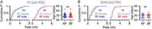
Optogenetic stimulation of parvalbumin- and somatostatin-expressing interneurons (PVs and SOMs) triggers inhibitory postsynaptic currents (IPSCs) in both regular- and burst-firing (RFs and BFs) subicular pyramidal cells.
The amplitude of optogenetically evoked IPSCs from PVs (PV-opto IPSCs) is larger in BFs whereas IPSCs generated by the light activation of SOMs (SOM-opto IPSCs) are larger in RFs.
Both PV- and SOM-opto IPSCs critically depend on ω-agatoxin IVA-sensitive P/Q type voltage-gated calcium channels, whereas no major effects are observed following exposure to ω-conotoxin GVIA, suggesting no significant involvement of N-type channels.
The amplitude of both PV- and SOM-opto IPSCs is reduced by the probable pharmacological activation of presynaptic μ opioid receptors, with a faster kinetics of the effect observed in PV-opto IPSCs from RFs vs. BFs, but not in SOM-opto IPSCs.
These results help us understand the complex interactions between different layers of diversity regulating GABAergic input onto subicular microcircuits.
期刊介绍:
The Journal of Physiology publishes full-length original Research Papers and Techniques for Physiology, which are short papers aimed at disseminating new techniques for physiological research. Articles solicited by the Editorial Board include Perspectives, Symposium Reports and Topical Reviews, which highlight areas of special physiological interest. CrossTalk articles are short editorial-style invited articles framing a debate between experts in the field on controversial topics. Letters to the Editor and Journal Club articles are also published. All categories of papers are subjected to peer reivew.
The Journal of Physiology welcomes submitted research papers in all areas of physiology. Authors should present original work that illustrates new physiological principles or mechanisms. Papers on work at the molecular level, at the level of the cell membrane, single cells, tissues or organs and on systems physiology are all acceptable. Theoretical papers and papers that use computational models to further our understanding of physiological processes will be considered if based on experimentally derived data and if the hypothesis advanced is directly amenable to experimental testing. While emphasis is on human and mammalian physiology, work on lower vertebrate or invertebrate preparations may be suitable if it furthers the understanding of the functioning of other organisms including mammals.


 分享
分享
 求助内容:
求助内容: 应助结果提醒方式:
 扫码关注我们
