{"title":"光谱双载体计算机断层扫描对肺腺癌 I 期 PD-L1 表达的预测价值:新型提名图的开发与验证。","authors":"Tong Wang, Zheng Fan, Yong Yue, Xiaomei Lu, Xiaoxu Deng, Yang Hou","doi":"10.21037/qims-24-15","DOIUrl":null,"url":null,"abstract":"<p><strong>Background: </strong>Programmed death ligand-1 (<i>PD-L1</i>) expression serves a predictive biomarker for the efficacy of immune checkpoint inhibitors (ICIs) in the treatment of patients with early-stage lung adenocarcinoma (LA). However, only a limited number of studies have explored the relationship between <i>PD-L1</i> expression and spectral dual-layer detector-based computed tomography (SDCT) quantification, qualitative parameters, and clinical biomarkers. Therefore, this study was conducted to clarify this relationship in stage I LA and to develop a nomogram to assist in preoperative individualized identification of <i>PD-L1</i>-positive expression.</p><p><strong>Methods: </strong>We analyzed SDCT parameters and <i>PD-L1</i> expression in patients diagnosed with invasive nonmucinous LA through postoperative pathology. Patients were categorized into <i>PD-L1</i>-positive and <i>PD-L1</i>-negative expression groups based on a threshold of 1%. A retrospective set (N=356) was used to develop and internally validate the radiological and biomarker features collected from predictive models. Univariate analysis was employed to reduce dimensionality, and logistic regression was used to establish a nomogram for predicting <i>PD-L1</i> expression. The predictive performance of the model was evaluated using receiver operating characteristic (ROC) curves, and external validation was performed in an independent set (N=80).</p><p><strong>Results: </strong>The proportions of solid components and pleural indentations were higher in the <i>PD-L1</i>-positive group, as indicated by the computed tomography (CT) value, CT at 40 keV (CT40keV; a/v), electron density (ED; a/v), and thymidine kinase 1 (TK1) exhibiting a positive correlation with <i>PD-L1</i> expression. In contrast, the effective atomic number (Zeff; a/v) showed a negative correlation with <i>PD-L1</i> expression [r=-0.4266 (Zeff.a), -0.1131 (Zeff.v); P<0.05]. After univariate analysis, 18 parameters were found to be associated with <i>PD-L1</i> expression. Multiple regression analysis was performed on significant parameters with an area under the curve (AUC) >0.6, and CT value [AUC =0.627; odds ratio (OR) =0.993; P=0.033], CT40keV.a (AUC =0.642; OR =1.006; P=0.025), arterial Zeff (Zeff.a) (AUC =0.756; OR =0.102; P<0.001), arterial ED (ED.a) (AUC =0.641; OR =1.158, P<0.001), venous ED (ED.v) (AUC =0.607; OR =0.864; P<0.001), TK1 (AUC =0.601; OR =1.245; P=0.026), and diameter of solid components (Dsolid) (AUC =0.632; OR =1.058; P=0.04) were found to be independent risk factors for PD-L1 expression in stage I LA. These seven predictive factors were integrated into the development of an SDCT parameter-clinical nomogram, which demonstrated satisfactory discrimination ability in the training set [AUC =0.853; 95% confidence interval (CI): 0.76-0.947], internal validation set (AUC =0.824; 95% CI: 0.775-0.874), and external validation set (AUC =0.825; 95% CI: 0.733-0.918). Decision curve analyses also revealed the highest net benefit for the nomogram across a broad threshold probability range (20-80%), with a clinical impact curve (CIC) indicating its clinical validity. Comparisons with other models demonstrated the superior discriminatory accuracy of the nomogram over any individual variable (all P values <0.05).</p><p><strong>Conclusions: </strong>Quantitative parameters derived from SDCT demonstrated the ability to predict for <i>PD-L1</i> expression in early-stage LA, with Zeff.a being notably effective. The nomogram established in combination with TK1 showed excellent predictive performance and good calibration. This approach may facilitate the improved noninvasive prediction of <i>PD-L1</i> expression.</p>","PeriodicalId":54267,"journal":{"name":"Quantitative Imaging in Medicine and Surgery","volume":"14 8","pages":"5983-6001"},"PeriodicalIF":2.3000,"publicationDate":"2024-08-01","publicationTypes":"Journal Article","fieldsOfStudy":null,"isOpenAccess":false,"openAccessPdf":"https://www.ncbi.nlm.nih.gov/pmc/articles/PMC11320513/pdf/","citationCount":"0","resultStr":"{\"title\":\"Predictive value of spectral dual-detector computed tomography for <i>PD-L1</i> expression in stage I lung adenocarcinoma: development and validation of a novel nomogram.\",\"authors\":\"Tong Wang, Zheng Fan, Yong Yue, Xiaomei Lu, Xiaoxu Deng, Yang Hou\",\"doi\":\"10.21037/qims-24-15\",\"DOIUrl\":null,\"url\":null,\"abstract\":\"<p><strong>Background: </strong>Programmed death ligand-1 (<i>PD-L1</i>) expression serves a predictive biomarker for the efficacy of immune checkpoint inhibitors (ICIs) in the treatment of patients with early-stage lung adenocarcinoma (LA). However, only a limited number of studies have explored the relationship between <i>PD-L1</i> expression and spectral dual-layer detector-based computed tomography (SDCT) quantification, qualitative parameters, and clinical biomarkers. Therefore, this study was conducted to clarify this relationship in stage I LA and to develop a nomogram to assist in preoperative individualized identification of <i>PD-L1</i>-positive expression.</p><p><strong>Methods: </strong>We analyzed SDCT parameters and <i>PD-L1</i> expression in patients diagnosed with invasive nonmucinous LA through postoperative pathology. Patients were categorized into <i>PD-L1</i>-positive and <i>PD-L1</i>-negative expression groups based on a threshold of 1%. A retrospective set (N=356) was used to develop and internally validate the radiological and biomarker features collected from predictive models. Univariate analysis was employed to reduce dimensionality, and logistic regression was used to establish a nomogram for predicting <i>PD-L1</i> expression. The predictive performance of the model was evaluated using receiver operating characteristic (ROC) curves, and external validation was performed in an independent set (N=80).</p><p><strong>Results: </strong>The proportions of solid components and pleural indentations were higher in the <i>PD-L1</i>-positive group, as indicated by the computed tomography (CT) value, CT at 40 keV (CT40keV; a/v), electron density (ED; a/v), and thymidine kinase 1 (TK1) exhibiting a positive correlation with <i>PD-L1</i> expression. In contrast, the effective atomic number (Zeff; a/v) showed a negative correlation with <i>PD-L1</i> expression [r=-0.4266 (Zeff.a), -0.1131 (Zeff.v); P<0.05]. After univariate analysis, 18 parameters were found to be associated with <i>PD-L1</i> expression. Multiple regression analysis was performed on significant parameters with an area under the curve (AUC) >0.6, and CT value [AUC =0.627; odds ratio (OR) =0.993; P=0.033], CT40keV.a (AUC =0.642; OR =1.006; P=0.025), arterial Zeff (Zeff.a) (AUC =0.756; OR =0.102; P<0.001), arterial ED (ED.a) (AUC =0.641; OR =1.158, P<0.001), venous ED (ED.v) (AUC =0.607; OR =0.864; P<0.001), TK1 (AUC =0.601; OR =1.245; P=0.026), and diameter of solid components (Dsolid) (AUC =0.632; OR =1.058; P=0.04) were found to be independent risk factors for PD-L1 expression in stage I LA. These seven predictive factors were integrated into the development of an SDCT parameter-clinical nomogram, which demonstrated satisfactory discrimination ability in the training set [AUC =0.853; 95% confidence interval (CI): 0.76-0.947], internal validation set (AUC =0.824; 95% CI: 0.775-0.874), and external validation set (AUC =0.825; 95% CI: 0.733-0.918). Decision curve analyses also revealed the highest net benefit for the nomogram across a broad threshold probability range (20-80%), with a clinical impact curve (CIC) indicating its clinical validity. Comparisons with other models demonstrated the superior discriminatory accuracy of the nomogram over any individual variable (all P values <0.05).</p><p><strong>Conclusions: </strong>Quantitative parameters derived from SDCT demonstrated the ability to predict for <i>PD-L1</i> expression in early-stage LA, with Zeff.a being notably effective. The nomogram established in combination with TK1 showed excellent predictive performance and good calibration. This approach may facilitate the improved noninvasive prediction of <i>PD-L1</i> expression.</p>\",\"PeriodicalId\":54267,\"journal\":{\"name\":\"Quantitative Imaging in Medicine and Surgery\",\"volume\":\"14 8\",\"pages\":\"5983-6001\"},\"PeriodicalIF\":2.3000,\"publicationDate\":\"2024-08-01\",\"publicationTypes\":\"Journal Article\",\"fieldsOfStudy\":null,\"isOpenAccess\":false,\"openAccessPdf\":\"https://www.ncbi.nlm.nih.gov/pmc/articles/PMC11320513/pdf/\",\"citationCount\":\"0\",\"resultStr\":null,\"platform\":\"Semanticscholar\",\"paperid\":null,\"PeriodicalName\":\"Quantitative Imaging in Medicine and Surgery\",\"FirstCategoryId\":\"3\",\"ListUrlMain\":\"https://doi.org/10.21037/qims-24-15\",\"RegionNum\":2,\"RegionCategory\":\"医学\",\"ArticlePicture\":[],\"TitleCN\":null,\"AbstractTextCN\":null,\"PMCID\":null,\"EPubDate\":\"2024/7/24 0:00:00\",\"PubModel\":\"Epub\",\"JCR\":\"Q2\",\"JCRName\":\"RADIOLOGY, NUCLEAR MEDICINE & MEDICAL IMAGING\",\"Score\":null,\"Total\":0}","platform":"Semanticscholar","paperid":null,"PeriodicalName":"Quantitative Imaging in Medicine and Surgery","FirstCategoryId":"3","ListUrlMain":"https://doi.org/10.21037/qims-24-15","RegionNum":2,"RegionCategory":"医学","ArticlePicture":[],"TitleCN":null,"AbstractTextCN":null,"PMCID":null,"EPubDate":"2024/7/24 0:00:00","PubModel":"Epub","JCR":"Q2","JCRName":"RADIOLOGY, NUCLEAR MEDICINE & MEDICAL IMAGING","Score":null,"Total":0}
Predictive value of spectral dual-detector computed tomography for PD-L1 expression in stage I lung adenocarcinoma: development and validation of a novel nomogram.
Background: Programmed death ligand-1 (PD-L1) expression serves a predictive biomarker for the efficacy of immune checkpoint inhibitors (ICIs) in the treatment of patients with early-stage lung adenocarcinoma (LA). However, only a limited number of studies have explored the relationship between PD-L1 expression and spectral dual-layer detector-based computed tomography (SDCT) quantification, qualitative parameters, and clinical biomarkers. Therefore, this study was conducted to clarify this relationship in stage I LA and to develop a nomogram to assist in preoperative individualized identification of PD-L1-positive expression.
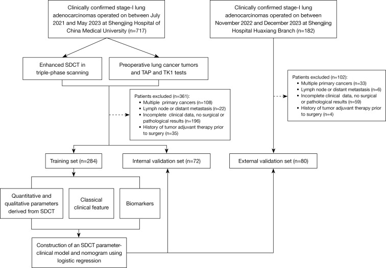
Methods: We analyzed SDCT parameters and PD-L1 expression in patients diagnosed with invasive nonmucinous LA through postoperative pathology. Patients were categorized into PD-L1-positive and PD-L1-negative expression groups based on a threshold of 1%. A retrospective set (N=356) was used to develop and internally validate the radiological and biomarker features collected from predictive models. Univariate analysis was employed to reduce dimensionality, and logistic regression was used to establish a nomogram for predicting PD-L1 expression. The predictive performance of the model was evaluated using receiver operating characteristic (ROC) curves, and external validation was performed in an independent set (N=80).
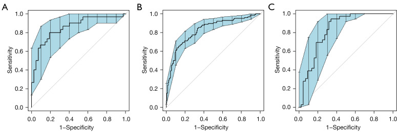
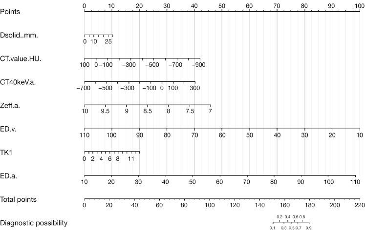
Results: The proportions of solid components and pleural indentations were higher in the PD-L1-positive group, as indicated by the computed tomography (CT) value, CT at 40 keV (CT40keV; a/v), electron density (ED; a/v), and thymidine kinase 1 (TK1) exhibiting a positive correlation with PD-L1 expression. In contrast, the effective atomic number (Zeff; a/v) showed a negative correlation with PD-L1 expression [r=-0.4266 (Zeff.a), -0.1131 (Zeff.v); P<0.05]. After univariate analysis, 18 parameters were found to be associated with PD-L1 expression. Multiple regression analysis was performed on significant parameters with an area under the curve (AUC) >0.6, and CT value [AUC =0.627; odds ratio (OR) =0.993; P=0.033], CT40keV.a (AUC =0.642; OR =1.006; P=0.025), arterial Zeff (Zeff.a) (AUC =0.756; OR =0.102; P<0.001), arterial ED (ED.a) (AUC =0.641; OR =1.158, P<0.001), venous ED (ED.v) (AUC =0.607; OR =0.864; P<0.001), TK1 (AUC =0.601; OR =1.245; P=0.026), and diameter of solid components (Dsolid) (AUC =0.632; OR =1.058; P=0.04) were found to be independent risk factors for PD-L1 expression in stage I LA. These seven predictive factors were integrated into the development of an SDCT parameter-clinical nomogram, which demonstrated satisfactory discrimination ability in the training set [AUC =0.853; 95% confidence interval (CI): 0.76-0.947], internal validation set (AUC =0.824; 95% CI: 0.775-0.874), and external validation set (AUC =0.825; 95% CI: 0.733-0.918). Decision curve analyses also revealed the highest net benefit for the nomogram across a broad threshold probability range (20-80%), with a clinical impact curve (CIC) indicating its clinical validity. Comparisons with other models demonstrated the superior discriminatory accuracy of the nomogram over any individual variable (all P values <0.05).
Conclusions: Quantitative parameters derived from SDCT demonstrated the ability to predict for PD-L1 expression in early-stage LA, with Zeff.a being notably effective. The nomogram established in combination with TK1 showed excellent predictive performance and good calibration. This approach may facilitate the improved noninvasive prediction of PD-L1 expression.


 分享
分享
 求助内容:
求助内容: 应助结果提醒方式:
 扫码关注我们
