{"title":"单个肌纤维肉瘤的磁共振成像、组织病理学和 Ki-67 标记指数结果比较:病例报告。","authors":"Ryusuke Tsujimura, Kenta Uto, Noriyuki Nakano, Yusuke Sato, Yuko Kobashi, Toshio Kojima, Hiroyuki Hao","doi":"10.1186/s13256-024-04693-y","DOIUrl":null,"url":null,"abstract":"<p><strong>Background: </strong>Myxofibrosarcoma is a myxoid soft tissue sarcoma showing T2 high intensity on magnetic resonance imaging. However, myxofibrosarcoma is a heterogeneous sarcoma with both myxoid and cellular portions. Magnetic resonance imaging findings were obtained MRI findings for comparison with histological and Ki-67 immunohistochemical features, in different portions of one myxofibrosarcoma.</p><p><strong>Case presentation: </strong>Magnetic resonance imaging observations were compared with gross pathological and microscopic findings of a myxofibrosarcoma from a 50-year-old Japanese female. The Ki-67 labeling indices of different portions of the tumor, that is, the myxoid, cellular, and histologically confirmed infiltrative margin portions (pathological tail sign), were compared. The T2 low intensity area was more cellular than the T2 high intensity area, while the cellular portion had a significantly higher Ki-67 index than the myxoid portion (p = 0.0313). The portions with the pathological tail sign had a significantly higher Ki-67 labeling index than those without this sign (p = 0.0313).</p><p><strong>Conclusions: </strong>More cellular portions of a myxofibrosarcoma correspond to more areas of the tumor showing aggressive features. Furthermore, our data also support the hypothesis of high aggressiveness being associated with the pathological tail sign in myxofibrosarcoma. To our knowledge, this is the first case report to describe comparisons among the imaging findings, histological features, and Ki-67 immunohistochemistry results for different portions of one myxofibrosarcoma.</p>","PeriodicalId":16236,"journal":{"name":"Journal of Medical Case Reports","volume":"18 1","pages":"373"},"PeriodicalIF":0.8000,"publicationDate":"2024-08-16","publicationTypes":"Journal Article","fieldsOfStudy":null,"isOpenAccess":false,"openAccessPdf":"https://www.ncbi.nlm.nih.gov/pmc/articles/PMC11328375/pdf/","citationCount":"0","resultStr":"{\"title\":\"Comparisons of magnetic resonance imaging, histopathological and Ki-67 labeling index findings in a single myxofibrosarcoma: a case report.\",\"authors\":\"Ryusuke Tsujimura, Kenta Uto, Noriyuki Nakano, Yusuke Sato, Yuko Kobashi, Toshio Kojima, Hiroyuki Hao\",\"doi\":\"10.1186/s13256-024-04693-y\",\"DOIUrl\":null,\"url\":null,\"abstract\":\"<p><strong>Background: </strong>Myxofibrosarcoma is a myxoid soft tissue sarcoma showing T2 high intensity on magnetic resonance imaging. However, myxofibrosarcoma is a heterogeneous sarcoma with both myxoid and cellular portions. Magnetic resonance imaging findings were obtained MRI findings for comparison with histological and Ki-67 immunohistochemical features, in different portions of one myxofibrosarcoma.</p><p><strong>Case presentation: </strong>Magnetic resonance imaging observations were compared with gross pathological and microscopic findings of a myxofibrosarcoma from a 50-year-old Japanese female. The Ki-67 labeling indices of different portions of the tumor, that is, the myxoid, cellular, and histologically confirmed infiltrative margin portions (pathological tail sign), were compared. The T2 low intensity area was more cellular than the T2 high intensity area, while the cellular portion had a significantly higher Ki-67 index than the myxoid portion (p = 0.0313). The portions with the pathological tail sign had a significantly higher Ki-67 labeling index than those without this sign (p = 0.0313).</p><p><strong>Conclusions: </strong>More cellular portions of a myxofibrosarcoma correspond to more areas of the tumor showing aggressive features. Furthermore, our data also support the hypothesis of high aggressiveness being associated with the pathological tail sign in myxofibrosarcoma. To our knowledge, this is the first case report to describe comparisons among the imaging findings, histological features, and Ki-67 immunohistochemistry results for different portions of one myxofibrosarcoma.</p>\",\"PeriodicalId\":16236,\"journal\":{\"name\":\"Journal of Medical Case Reports\",\"volume\":\"18 1\",\"pages\":\"373\"},\"PeriodicalIF\":0.8000,\"publicationDate\":\"2024-08-16\",\"publicationTypes\":\"Journal Article\",\"fieldsOfStudy\":null,\"isOpenAccess\":false,\"openAccessPdf\":\"https://www.ncbi.nlm.nih.gov/pmc/articles/PMC11328375/pdf/\",\"citationCount\":\"0\",\"resultStr\":null,\"platform\":\"Semanticscholar\",\"paperid\":null,\"PeriodicalName\":\"Journal of Medical Case Reports\",\"FirstCategoryId\":\"1085\",\"ListUrlMain\":\"https://doi.org/10.1186/s13256-024-04693-y\",\"RegionNum\":0,\"RegionCategory\":null,\"ArticlePicture\":[],\"TitleCN\":null,\"AbstractTextCN\":null,\"PMCID\":null,\"EPubDate\":\"\",\"PubModel\":\"\",\"JCR\":\"Q3\",\"JCRName\":\"MEDICINE, GENERAL & INTERNAL\",\"Score\":null,\"Total\":0}","platform":"Semanticscholar","paperid":null,"PeriodicalName":"Journal of Medical Case Reports","FirstCategoryId":"1085","ListUrlMain":"https://doi.org/10.1186/s13256-024-04693-y","RegionNum":0,"RegionCategory":null,"ArticlePicture":[],"TitleCN":null,"AbstractTextCN":null,"PMCID":null,"EPubDate":"","PubModel":"","JCR":"Q3","JCRName":"MEDICINE, GENERAL & INTERNAL","Score":null,"Total":0}
Comparisons of magnetic resonance imaging, histopathological and Ki-67 labeling index findings in a single myxofibrosarcoma: a case report.
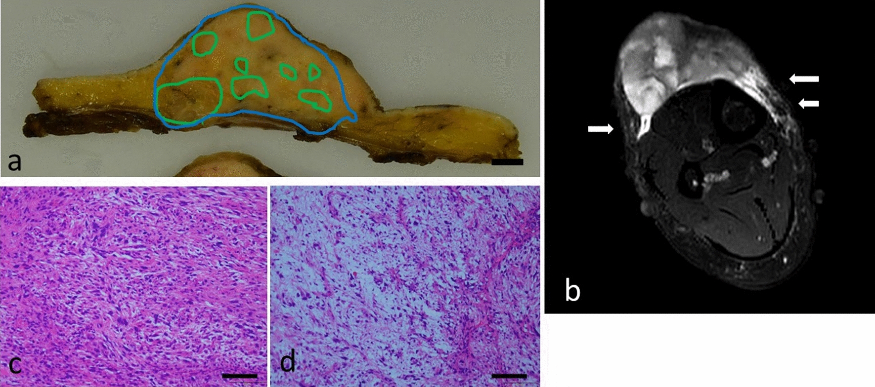
Background: Myxofibrosarcoma is a myxoid soft tissue sarcoma showing T2 high intensity on magnetic resonance imaging. However, myxofibrosarcoma is a heterogeneous sarcoma with both myxoid and cellular portions. Magnetic resonance imaging findings were obtained MRI findings for comparison with histological and Ki-67 immunohistochemical features, in different portions of one myxofibrosarcoma.
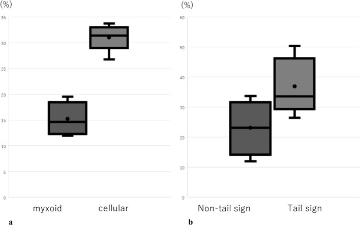
Case presentation: Magnetic resonance imaging observations were compared with gross pathological and microscopic findings of a myxofibrosarcoma from a 50-year-old Japanese female. The Ki-67 labeling indices of different portions of the tumor, that is, the myxoid, cellular, and histologically confirmed infiltrative margin portions (pathological tail sign), were compared. The T2 low intensity area was more cellular than the T2 high intensity area, while the cellular portion had a significantly higher Ki-67 index than the myxoid portion (p = 0.0313). The portions with the pathological tail sign had a significantly higher Ki-67 labeling index than those without this sign (p = 0.0313).
Conclusions: More cellular portions of a myxofibrosarcoma correspond to more areas of the tumor showing aggressive features. Furthermore, our data also support the hypothesis of high aggressiveness being associated with the pathological tail sign in myxofibrosarcoma. To our knowledge, this is the first case report to describe comparisons among the imaging findings, histological features, and Ki-67 immunohistochemistry results for different portions of one myxofibrosarcoma.
期刊介绍:
JMCR is an open access, peer-reviewed online journal that will consider any original case report that expands the field of general medical knowledge. Reports should show one of the following: 1. Unreported or unusual side effects or adverse interactions involving medications 2. Unexpected or unusual presentations of a disease 3. New associations or variations in disease processes 4. Presentations, diagnoses and/or management of new and emerging diseases 5. An unexpected association between diseases or symptoms 6. An unexpected event in the course of observing or treating a patient 7. Findings that shed new light on the possible pathogenesis of a disease or an adverse effect


 分享
分享
 求助内容:
求助内容: 应助结果提醒方式:
 扫码关注我们
