Tingting Yang , Jian Li , Xinyu Cheng , Qiuyuan Lu , Zara Farooq , Ying Fu , Sijia Lv , Weiwei Nan , Boming Yu , Jingjing Duan , Yuting Zhang , Yang Fu , Haihai Jiang , Peter J McCormick , Yanyan Li , Jin Zhang
{"title":"利用低温电子显微镜对人类 C5a-C5aR1 复合物进行结构分析。","authors":"Tingting Yang , Jian Li , Xinyu Cheng , Qiuyuan Lu , Zara Farooq , Ying Fu , Sijia Lv , Weiwei Nan , Boming Yu , Jingjing Duan , Yuting Zhang , Yang Fu , Haihai Jiang , Peter J McCormick , Yanyan Li , Jin Zhang","doi":"10.1016/j.jsb.2024.108117","DOIUrl":null,"url":null,"abstract":"<div><p>The complement system is a complex network of proteins that plays a crucial role in the innate immune response. One important component of this system is the C5a-C5aR1 complex, which is critical in the recruitment and activation of immune cells. In-depth investigation of the activation mechanism as well as biased signaling of the C5a-C5aR1 system will facilitate the elucidation of C5a-mediated pathophysiology. In this study, we determined the structure of C5a-C5aR1-Gi complex at a high resolution of 3 Å using cryo-electron microscopy (Cryo-EM). Our results revealed the binding site of C5a, which consists of a polar recognition region on the extracellular side and an amphipathic pocket within the transmembrane domain. Furthermore, we found that C5a binding induces conformational changes of C5aR1, which subsequently leads to the activation of G protein signaling pathways. Notably, a key residue (M265) located on transmembrane helix 6 (TM6) was identified to play a crucial role in regulating the recruitment of β-arrestin driven by C5a. This study provides more information about the structure and function of the human C5a-C5aR1 complex, which is essential for the proper functioning of the complement system. The findings of this study can also provide a foundation for the design of new pharmaceuticals targeting this receptor with bias or specificity.</p></div>","PeriodicalId":17074,"journal":{"name":"Journal of structural biology","volume":"216 3","pages":"Article 108117"},"PeriodicalIF":2.7000,"publicationDate":"2024-09-01","publicationTypes":"Journal Article","fieldsOfStudy":null,"isOpenAccess":false,"openAccessPdf":"","citationCount":"0","resultStr":"{\"title\":\"Structural analysis of the human C5a-C5aR1 complex using cryo-electron microscopy\",\"authors\":\"Tingting Yang , Jian Li , Xinyu Cheng , Qiuyuan Lu , Zara Farooq , Ying Fu , Sijia Lv , Weiwei Nan , Boming Yu , Jingjing Duan , Yuting Zhang , Yang Fu , Haihai Jiang , Peter J McCormick , Yanyan Li , Jin Zhang\",\"doi\":\"10.1016/j.jsb.2024.108117\",\"DOIUrl\":null,\"url\":null,\"abstract\":\"<div><p>The complement system is a complex network of proteins that plays a crucial role in the innate immune response. One important component of this system is the C5a-C5aR1 complex, which is critical in the recruitment and activation of immune cells. In-depth investigation of the activation mechanism as well as biased signaling of the C5a-C5aR1 system will facilitate the elucidation of C5a-mediated pathophysiology. In this study, we determined the structure of C5a-C5aR1-Gi complex at a high resolution of 3 Å using cryo-electron microscopy (Cryo-EM). Our results revealed the binding site of C5a, which consists of a polar recognition region on the extracellular side and an amphipathic pocket within the transmembrane domain. Furthermore, we found that C5a binding induces conformational changes of C5aR1, which subsequently leads to the activation of G protein signaling pathways. Notably, a key residue (M265) located on transmembrane helix 6 (TM6) was identified to play a crucial role in regulating the recruitment of β-arrestin driven by C5a. This study provides more information about the structure and function of the human C5a-C5aR1 complex, which is essential for the proper functioning of the complement system. The findings of this study can also provide a foundation for the design of new pharmaceuticals targeting this receptor with bias or specificity.</p></div>\",\"PeriodicalId\":17074,\"journal\":{\"name\":\"Journal of structural biology\",\"volume\":\"216 3\",\"pages\":\"Article 108117\"},\"PeriodicalIF\":2.7000,\"publicationDate\":\"2024-09-01\",\"publicationTypes\":\"Journal Article\",\"fieldsOfStudy\":null,\"isOpenAccess\":false,\"openAccessPdf\":\"\",\"citationCount\":\"0\",\"resultStr\":null,\"platform\":\"Semanticscholar\",\"paperid\":null,\"PeriodicalName\":\"Journal of structural biology\",\"FirstCategoryId\":\"99\",\"ListUrlMain\":\"https://www.sciencedirect.com/science/article/pii/S1047847724000571\",\"RegionNum\":3,\"RegionCategory\":\"生物学\",\"ArticlePicture\":[],\"TitleCN\":null,\"AbstractTextCN\":null,\"PMCID\":null,\"EPubDate\":\"2024/8/15 0:00:00\",\"PubModel\":\"Epub\",\"JCR\":\"Q3\",\"JCRName\":\"BIOCHEMISTRY & MOLECULAR BIOLOGY\",\"Score\":null,\"Total\":0}","platform":"Semanticscholar","paperid":null,"PeriodicalName":"Journal of structural biology","FirstCategoryId":"99","ListUrlMain":"https://www.sciencedirect.com/science/article/pii/S1047847724000571","RegionNum":3,"RegionCategory":"生物学","ArticlePicture":[],"TitleCN":null,"AbstractTextCN":null,"PMCID":null,"EPubDate":"2024/8/15 0:00:00","PubModel":"Epub","JCR":"Q3","JCRName":"BIOCHEMISTRY & MOLECULAR BIOLOGY","Score":null,"Total":0}
Structural analysis of the human C5a-C5aR1 complex using cryo-electron microscopy
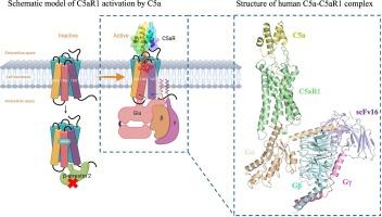
The complement system is a complex network of proteins that plays a crucial role in the innate immune response. One important component of this system is the C5a-C5aR1 complex, which is critical in the recruitment and activation of immune cells. In-depth investigation of the activation mechanism as well as biased signaling of the C5a-C5aR1 system will facilitate the elucidation of C5a-mediated pathophysiology. In this study, we determined the structure of C5a-C5aR1-Gi complex at a high resolution of 3 Å using cryo-electron microscopy (Cryo-EM). Our results revealed the binding site of C5a, which consists of a polar recognition region on the extracellular side and an amphipathic pocket within the transmembrane domain. Furthermore, we found that C5a binding induces conformational changes of C5aR1, which subsequently leads to the activation of G protein signaling pathways. Notably, a key residue (M265) located on transmembrane helix 6 (TM6) was identified to play a crucial role in regulating the recruitment of β-arrestin driven by C5a. This study provides more information about the structure and function of the human C5a-C5aR1 complex, which is essential for the proper functioning of the complement system. The findings of this study can also provide a foundation for the design of new pharmaceuticals targeting this receptor with bias or specificity.
期刊介绍:
Journal of Structural Biology (JSB) has an open access mirror journal, the Journal of Structural Biology: X (JSBX), sharing the same aims and scope, editorial team, submission system and rigorous peer review. Since both journals share the same editorial system, you may submit your manuscript via either journal homepage. You will be prompted during submission (and revision) to choose in which to publish your article. The editors and reviewers are not aware of the choice you made until the article has been published online. JSB and JSBX publish papers dealing with the structural analysis of living material at every level of organization by all methods that lead to an understanding of biological function in terms of molecular and supermolecular structure.
Techniques covered include:
• Light microscopy including confocal microscopy
• All types of electron microscopy
• X-ray diffraction
• Nuclear magnetic resonance
• Scanning force microscopy, scanning probe microscopy, and tunneling microscopy
• Digital image processing
• Computational insights into structure


 分享
分享
 求助内容:
求助内容: 应助结果提醒方式:
 扫码关注我们
