Fahad N Altamimi, AlTheyab Fatemah, Mariam Al-Amro, Ali Al Montasher, Sara Al Otaibi, Fida Al Muhawas
{"title":"人工耳蜗植入:耳蜗直径小可能表明异常程度。","authors":"Fahad N Altamimi, AlTheyab Fatemah, Mariam Al-Amro, Ali Al Montasher, Sara Al Otaibi, Fida Al Muhawas","doi":"10.5152/iao.2024.231191","DOIUrl":null,"url":null,"abstract":"<p><p>Cochlear size variation was first reported in 1884, and since then, there have been various reports confirming the same. Yet, there is no single report that has displayed the wide variations in the cochlear size in a single layout capturing the cochlea in the oblique coronal view/ cochlear view. Basal turn diameter (A-value) was measured in the oblique coronal plane using the OTOPLAN® otological preplanning tool in 104 computed tomography (CT) scans of the temporal bones of cochlear implant (CI) recipients in a tertiary CI center. All CT scans with an image resolution of at least 0.5 mm and identified as having cochleae with normal anatomy were included in this study. A 3-dimensional (3D) segmentation was performed using the 3D slicer and visualized to evaluate the impact of cochlear size on the number of turns studied. The A-value was found to vary between 7.3 mm and 10.4 mm among the studied patients. Three-dimensional segmentation of the inner ear revealed only 2 turns of the cochlea in 4 ears, with A-values of 7.3, 8.8, 7.8, and 7.7 mm. One ear had only 11 /2 turns of the cochlea, with an A-value of 7.9 mm. As a further advancement in the assessment of cochlear size as determined by the A-value, 3D segmentation of the complete inner ear provides a full picture of the number of cochlear turns. Three-dimensional segmentation of the entire inner ear could help improve the preoperative planning of CI surgery and have implications for electrode array selection. Cochlear size could be a predictor of the number of cochlear turns, even in cases that look normal from the radiological findings. The findings of this study could help in improving the preoperative planning for a more successful CI surgery by differentiating between the normal and abnormal cochlea.</p>","PeriodicalId":94238,"journal":{"name":"The journal of international advanced otology","volume":"20 2","pages":"108-112"},"PeriodicalIF":1.2000,"publicationDate":"2024-03-27","publicationTypes":"Journal Article","fieldsOfStudy":null,"isOpenAccess":false,"openAccessPdf":"https://www.ncbi.nlm.nih.gov/pmc/articles/PMC11114178/pdf/","citationCount":"0","resultStr":"{\"title\":\"Cochlear Implantation: Small Cochlear Diameter May Indicate Degree of Abnormality.\",\"authors\":\"Fahad N Altamimi, AlTheyab Fatemah, Mariam Al-Amro, Ali Al Montasher, Sara Al Otaibi, Fida Al Muhawas\",\"doi\":\"10.5152/iao.2024.231191\",\"DOIUrl\":null,\"url\":null,\"abstract\":\"<p><p>Cochlear size variation was first reported in 1884, and since then, there have been various reports confirming the same. Yet, there is no single report that has displayed the wide variations in the cochlear size in a single layout capturing the cochlea in the oblique coronal view/ cochlear view. Basal turn diameter (A-value) was measured in the oblique coronal plane using the OTOPLAN® otological preplanning tool in 104 computed tomography (CT) scans of the temporal bones of cochlear implant (CI) recipients in a tertiary CI center. All CT scans with an image resolution of at least 0.5 mm and identified as having cochleae with normal anatomy were included in this study. A 3-dimensional (3D) segmentation was performed using the 3D slicer and visualized to evaluate the impact of cochlear size on the number of turns studied. The A-value was found to vary between 7.3 mm and 10.4 mm among the studied patients. Three-dimensional segmentation of the inner ear revealed only 2 turns of the cochlea in 4 ears, with A-values of 7.3, 8.8, 7.8, and 7.7 mm. One ear had only 11 /2 turns of the cochlea, with an A-value of 7.9 mm. As a further advancement in the assessment of cochlear size as determined by the A-value, 3D segmentation of the complete inner ear provides a full picture of the number of cochlear turns. Three-dimensional segmentation of the entire inner ear could help improve the preoperative planning of CI surgery and have implications for electrode array selection. Cochlear size could be a predictor of the number of cochlear turns, even in cases that look normal from the radiological findings. The findings of this study could help in improving the preoperative planning for a more successful CI surgery by differentiating between the normal and abnormal cochlea.</p>\",\"PeriodicalId\":94238,\"journal\":{\"name\":\"The journal of international advanced otology\",\"volume\":\"20 2\",\"pages\":\"108-112\"},\"PeriodicalIF\":1.2000,\"publicationDate\":\"2024-03-27\",\"publicationTypes\":\"Journal Article\",\"fieldsOfStudy\":null,\"isOpenAccess\":false,\"openAccessPdf\":\"https://www.ncbi.nlm.nih.gov/pmc/articles/PMC11114178/pdf/\",\"citationCount\":\"0\",\"resultStr\":null,\"platform\":\"Semanticscholar\",\"paperid\":null,\"PeriodicalName\":\"The journal of international advanced otology\",\"FirstCategoryId\":\"1085\",\"ListUrlMain\":\"https://doi.org/10.5152/iao.2024.231191\",\"RegionNum\":0,\"RegionCategory\":null,\"ArticlePicture\":[],\"TitleCN\":null,\"AbstractTextCN\":null,\"PMCID\":null,\"EPubDate\":\"\",\"PubModel\":\"\",\"JCR\":\"\",\"JCRName\":\"\",\"Score\":null,\"Total\":0}","platform":"Semanticscholar","paperid":null,"PeriodicalName":"The journal of international advanced otology","FirstCategoryId":"1085","ListUrlMain":"https://doi.org/10.5152/iao.2024.231191","RegionNum":0,"RegionCategory":null,"ArticlePicture":[],"TitleCN":null,"AbstractTextCN":null,"PMCID":null,"EPubDate":"","PubModel":"","JCR":"","JCRName":"","Score":null,"Total":0}
Cochlear Implantation: Small Cochlear Diameter May Indicate Degree of Abnormality.
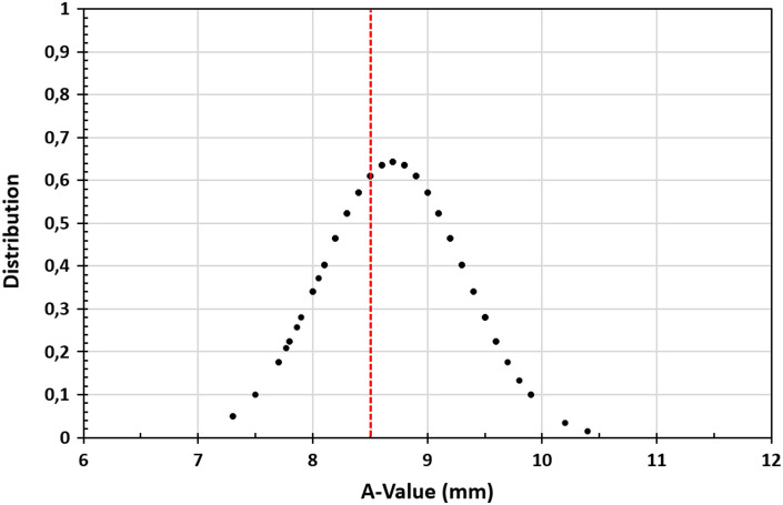
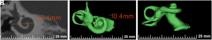
Cochlear size variation was first reported in 1884, and since then, there have been various reports confirming the same. Yet, there is no single report that has displayed the wide variations in the cochlear size in a single layout capturing the cochlea in the oblique coronal view/ cochlear view. Basal turn diameter (A-value) was measured in the oblique coronal plane using the OTOPLAN® otological preplanning tool in 104 computed tomography (CT) scans of the temporal bones of cochlear implant (CI) recipients in a tertiary CI center. All CT scans with an image resolution of at least 0.5 mm and identified as having cochleae with normal anatomy were included in this study. A 3-dimensional (3D) segmentation was performed using the 3D slicer and visualized to evaluate the impact of cochlear size on the number of turns studied. The A-value was found to vary between 7.3 mm and 10.4 mm among the studied patients. Three-dimensional segmentation of the inner ear revealed only 2 turns of the cochlea in 4 ears, with A-values of 7.3, 8.8, 7.8, and 7.7 mm. One ear had only 11 /2 turns of the cochlea, with an A-value of 7.9 mm. As a further advancement in the assessment of cochlear size as determined by the A-value, 3D segmentation of the complete inner ear provides a full picture of the number of cochlear turns. Three-dimensional segmentation of the entire inner ear could help improve the preoperative planning of CI surgery and have implications for electrode array selection. Cochlear size could be a predictor of the number of cochlear turns, even in cases that look normal from the radiological findings. The findings of this study could help in improving the preoperative planning for a more successful CI surgery by differentiating between the normal and abnormal cochlea.


 分享
分享

 求助内容:
求助内容: 应助结果提醒方式:
 扫码关注我们
